6MNI | pdb_00006mni

Structure of the tandem CACHE domain of PscC


Experimental Data Snapshot

  • Method: X-RAY DIFFRACTION
  • Resolution: 1.70 Å
  • R-Value Free: 
    0.200 (Depositor), 0.200 (DCC) 
  • R-Value Work: 
    0.162 (Depositor), 0.160 (DCC) 
  • R-Value Observed: 
    0.164 (Depositor) 

Starting Model: experimental
View more details

wwPDB Validation   3D Report Full Report


Ligand Structure Quality Assessment 

Created with Raphaël 2.3.0Worse 01 BetterLigand structure goodness of fit to experimental dataBest fitted PROClick on this verticalbar to view details

This is version 1.2 of the entry. See complete history


Literature

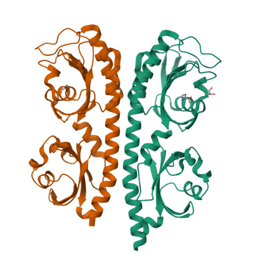
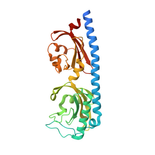
Structure of a double CACHE chemoreceptor ligand-binding domain from Pseudomonas syringae provides insights into the basis of proline recognition.

Ehrhardt, M.K.G.Gerth, M.L.Johnston, J.M.

(2021) Biochem Biophys Res Commun 549: 194-199

  • DOI: https://doi.org/10.1016/j.bbrc.2021.02.090
  • Primary Citation of Related Structures:  
    6MNI

  • PubMed Abstract: 

    Chemotaxis is the process of sensing chemical gradients and navigating towards favourable conditions. Bacterial chemotaxis is mediated by arrays of trans-membrane chemoreceptor proteins. The most common class of chemoreceptors have periplasmic ligand-binding domains (LBDs) that detect extracellular chemical signs and transduce these signals to the downstream chemotaxis machinery. The repertoire of chemoreceptor proteins in a bacterium determines the range of environmental signals to which it can respond. Pseudomonas syringae pv. actinidiae (Psa) is a plant pathogen which causes bacterial canker of kiwifruit (Actinidia sp.). Compared to many other bacteria, Psa has a large number of chemoreceptors encoded in its genome (43) and most of these remain uncharacterized. A previous study identified PscC as a potential chemoreceptor for l-proline and other amino acid ligands. Here, we have characterized the interaction of PscC-LBD with l-proline using a combination of isothermal titration calorimetry (ITC) and X-ray crystallography. ITC confirmed direct binding of l-proline to PscC-LBD with K D value of 5.0 μM. We determined the structure of PscC-LBD in complex with l-proline. Our structural analysis showed that PscC-LBD adopts similar double-CACHE fold to several other amino acid chemoreceptors. A comparison of the PscC-LDB to other dCACHE structures highlights residues in the binding cavity which contribute to its ligand specificity.


  • Organizational Affiliation

    Department of Biochemistry, University of Otago, Dunedin, New Zealand.


Macromolecules
Find similar proteins by:  (by identity cutoff)  |  3D Structure
Entity ID: 1
MoleculeChains Sequence LengthOrganismDetailsImage
Methyl-accepting chemotaxis protein
A, B
287Pseudomonas syringae pv. actinidiaeMutation(s): 0 
Gene Names: KPSA3_03559
Entity Groups  
Sequence Clusters30% Identity50% Identity70% Identity90% Identity95% Identity100% Identity
Sequence Annotations
Expand
  • Reference Sequence
Experimental Data & Validation

Experimental Data

  • Method: X-RAY DIFFRACTION
  • Resolution: 1.70 Å
  • R-Value Free:  0.200 (Depositor), 0.200 (DCC) 
  • R-Value Work:  0.162 (Depositor), 0.160 (DCC) 
  • R-Value Observed: 0.164 (Depositor) 
Space Group: P 1
Unit Cell:
Length ( Å )Angle ( ˚ )
a = 34.244α = 104.79
b = 53.916β = 91.2
c = 73.312γ = 92.71
Software Package:
Software NamePurpose
XDSdata reduction
Aimlessdata scaling
PHENIXrefinement
PDB_EXTRACTdata extraction
BALBESphasing

Structure Validation

View Full Validation Report


Ligand Structure Quality Assessment 

Created with Raphaël 2.3.0Worse 01 BetterLigand structure goodness of fit to experimental dataBest fitted PROClick on this verticalbar to view details

Entry History 

Deposition Data

Revision History  (Full details and data files)

  • Version 1.0: 2019-10-16
    Type: Initial release
  • Version 1.1: 2021-04-28
    Changes: Database references
  • Version 1.2: 2023-10-11
    Changes: Data collection, Database references, Refinement description