7B74 | pdb_00007b74

Chimeric Streptavidin With A Dimerization Domain For Artificial Transfer Hydrogenation


Experimental Data Snapshot

  • Method: X-RAY DIFFRACTION
  • Resolution: 1.85 Å
  • R-Value Free: 
    0.218 (Depositor), 0.230 (DCC) 
  • R-Value Work: 
    0.191 (Depositor), 0.200 (DCC) 

Starting Model: experimental
View more details

wwPDB Validation   3D Report Full Report


Ligand Structure Quality Assessment 

Created with Raphaël 2.3.0Worse 01 BetterLigand structure goodness of fit to experimental dataBest fitted 4IRClick on this verticalbar to view details

This is version 1.2 of the entry. See complete history


Literature

Spiers Memorial Lecture: Shielding the active site: a streptavidin superoxide-dismutase chimera as a host protein for asymmetric transfer hydrogenation.

Igareta, N.V.Tachibana, R.Spiess, D.C.Peterson, R.L.Ward, T.R.

(2023) Faraday Discuss 

  • DOI: https://doi.org/10.1039/d3fd00034f
  • Primary Citation of Related Structures:  
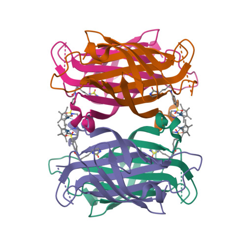
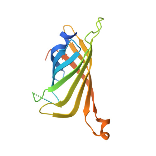
    7B74

  • PubMed Abstract: 

    By anchoring a metal cofactor within a host protein, so-called artificial metalloenzymes can be generated. Such hybrid catalysts combine the versatility of transition metals in catalyzing new-to-nature reactions with the power of genetic-engineering to evolve proteins. With the aim of gaining better control over second coordination-sphere interactions between a streptavidin host-protein (Sav) and a biotinylated cofactor, we engineered a hydrophobic dimerization domain, borrowed from superoxide dismutase C (SOD), on Sav's biotin-binding vestibule. The influence of the SOD dimerization domain (DD) on the performance of an asymmetric transfer hydrogenase (ATHase) resulting from anchoring a biotinylated Cp*Ir-cofactor - [Cp*Ir(biot- p -L)Cl] (1-Cl) - within Sav-SOD is reported herein. We show that, depending on the nature of the residue at position Sav S112, the introduction of the SOD DD on the biotin-binding vestibule leads to an inversion of configuration of the reduction product, as well as a fivefold increase in catalytic efficiency. The findings are rationalized by QM/MM calculations, combined with X-ray crystallography.


  • Organizational Affiliation

    Department of Chemistry, University of Basel, Mattenstrasse 24a, BPR 1096, Basel, CH-4058, Switzerland. thomas.ward@unibas.ch.


Macromolecules
Find similar proteins by:  (by identity cutoff)  |  3D Structure
Entity ID: 1
MoleculeChains Sequence LengthOrganismDetailsImage
Streptavidin,Superoxide dismutase [Cu-Zn],StreptavidinA [auth AAA],
B [auth BBB],
C [auth CCC],
D [auth DDD]
194Streptomyces avidiniiMycobacterium tuberculosis variant bovis AF2122/97Mutation(s): 1 
EC: 1.15.1.1
UniProt
Find proteins for P22629 (Streptomyces avidinii)
Explore P22629 
Go to UniProtKB:  P22629
Find proteins for P0A609 (Mycobacterium bovis (strain ATCC BAA-935 / AF2122/97))
Explore P0A609 
Go to UniProtKB:  P0A609
Entity Groups  
Sequence Clusters30% Identity50% Identity70% Identity90% Identity95% Identity100% Identity
UniProt GroupsP22629P0A609
Sequence Annotations
Expand
  • Reference Sequence
Experimental Data & Validation

Experimental Data

  • Method: X-RAY DIFFRACTION
  • Resolution: 1.85 Å
  • R-Value Free:  0.218 (Depositor), 0.230 (DCC) 
  • R-Value Work:  0.191 (Depositor), 0.200 (DCC) 
Space Group: P 1 21 1
Unit Cell:
Length ( Å )Angle ( ˚ )
a = 57.285α = 90
b = 57.307β = 94.616
c = 88.035γ = 90
Software Package:
Software NamePurpose
REFMACrefinement
REFMACrefinement
Aimlessdata reduction
XDSdata scaling
PHASERphasing

Structure Validation

View Full Validation Report


Ligand Structure Quality Assessment 

Created with Raphaël 2.3.0Worse 01 BetterLigand structure goodness of fit to experimental dataBest fitted 4IRClick on this verticalbar to view details

Entry History 

Deposition Data

Revision History  (Full details and data files)

  • Version 1.0: 2021-11-17
    Type: Initial release
  • Version 1.1: 2023-06-07
    Changes: Database references, Derived calculations
  • Version 1.2: 2024-02-07
    Changes: Data collection, Refinement description