7E3J

Crystal structure of SARS-CoV-2 RBD binding to dog ACE2


Experimental Data Snapshot

  • Method: X-RAY DIFFRACTION
  • Resolution: 2.99 Å
  • R-Value Free: 0.248 
  • R-Value Work: 0.227 
  • R-Value Observed: 0.228 

wwPDB Validation   3D Report Full Report


Ligand Structure Quality Assessment 


This is version 1.5 of the entry. See complete history


Literature

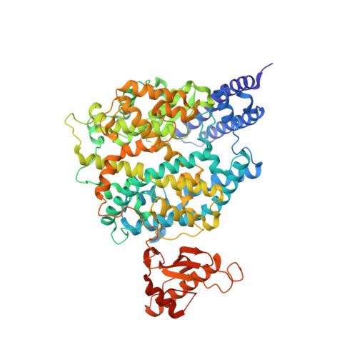
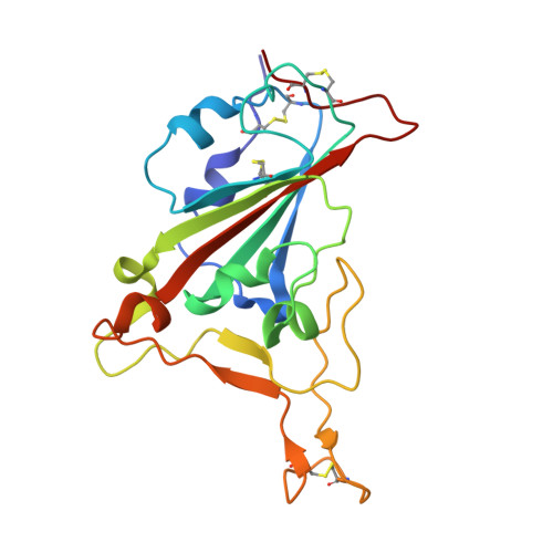
The molecular basis for SARS-CoV-2 binding to dog ACE2.

Zhang, Z.Zhang, Y.Liu, K.Li, Y.Lu, Q.Wang, Q.Zhang, Y.Wang, L.Liao, H.Zheng, A.Ma, S.Fan, Z.Li, H.Huang, W.Bi, Y.Zhao, X.Wang, Q.Gao, G.F.Xiao, H.Tong, Z.Qi, J.Sun, Y.

(2021) Nat Commun 12: 4195-4195

  • DOI: https://doi.org/10.1038/s41467-021-24326-y
  • Primary Citation of Related Structures:  
    7E3J

  • PubMed Abstract: 

    SARS-CoV-2 can infect many domestic animals, including dogs. Herein, we show that dog angiotensin-converting enzyme 2 (dACE2) can bind to the SARS-CoV-2 spike (S) protein receptor binding domain (RBD), and that both pseudotyped and authentic SARS-CoV-2 can infect dACE2-expressing cells. We solved the crystal structure of RBD in complex with dACE2 and found that the total number of contact residues, contact atoms, hydrogen bonds and salt bridges at the binding interface in this complex are slightly fewer than those in the complex of the RBD and human ACE2 (hACE2). This result is consistent with the fact that the binding affinity of RBD to dACE2 is lower than that of hACE2. We further show that a few important mutations in the RBD binding interface play a pivotal role in the binding affinity of RBD to both dACE2 and hACE2. Our work reveals a molecular basis for cross-species transmission and potential animal spread of SARS-CoV-2, and provides new clues to block the potential transmission chains of this virus.


  • Organizational Affiliation

    CAS Key Laboratory of Pathogenic Microbiology and Immunology, Institute of Microbiology, Chinese Academy of Sciences, Beijing, China.


Macromolecules
Find similar proteins by:  (by identity cutoff)  |  3D Structure
Entity ID: 1
MoleculeChains Sequence LengthOrganismDetailsImage
ACE2723Canis lupus familiarisMutation(s): 0 
Entity Groups  
Sequence Clusters30% Identity50% Identity70% Identity90% Identity95% Identity100% Identity
Sequence Annotations
Expand
  • Reference Sequence
Find similar proteins by:  (by identity cutoff)  |  3D Structure
Entity ID: 2
MoleculeChains Sequence LengthOrganismDetailsImage
Spike protein S1195Severe acute respiratory syndrome coronavirus 2Mutation(s): 0 
Gene Names: S2
UniProt
Find proteins for P0DTC2 (Severe acute respiratory syndrome coronavirus 2)
Explore P0DTC2 
Go to UniProtKB:  P0DTC2
Entity Groups  
Sequence Clusters30% Identity50% Identity70% Identity90% Identity95% Identity100% Identity
UniProt GroupP0DTC2
Sequence Annotations
Expand
  • Reference Sequence
Small Molecules
Experimental Data & Validation

Experimental Data

  • Method: X-RAY DIFFRACTION
  • Resolution: 2.99 Å
  • R-Value Free: 0.248 
  • R-Value Work: 0.227 
  • R-Value Observed: 0.228 
  • Space Group: I 4 2 2
Unit Cell:
Length ( Å )Angle ( ˚ )
a = 168.256α = 90
b = 168.256β = 90
c = 211.196γ = 90
Software Package:
Software NamePurpose
PHENIXrefinement
HKL-2000data reduction
HKL-2000data scaling
PHASERphasing

Structure Validation

View Full Validation Report


Ligand Structure Quality Assessment 


Entry History 

Deposition Data

Revision History  (Full details and data files)

  • Version 1.0: 2021-04-07
    Type: Initial release
  • Version 1.1: 2021-07-07
    Changes: Database references
  • Version 1.2: 2021-07-28
    Changes: Source and taxonomy
  • Version 1.3: 2021-08-25
    Changes: Database references, Structure summary
  • Version 1.4: 2021-10-13
    Changes: Database references
  • Version 1.5: 2023-11-29
    Changes: Data collection, Refinement description