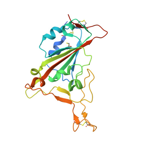
7EFP

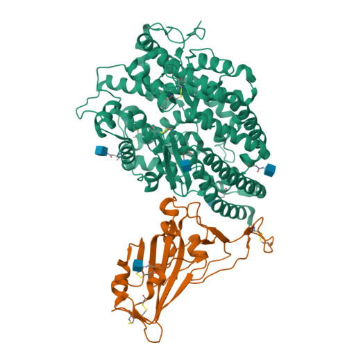
Structure of SARS-CoV-2 spike receptor-binding domain in complex with high affinity ACE2 mutant (S19W,N330Y)


Experimental Data Snapshot

  • Method: X-RAY DIFFRACTION
  • Resolution: 2.70 Å
  • R-Value Free: 0.224 
  • R-Value Work: 0.184 
  • R-Value Observed: 0.186 

Starting Model: experimental
View more details

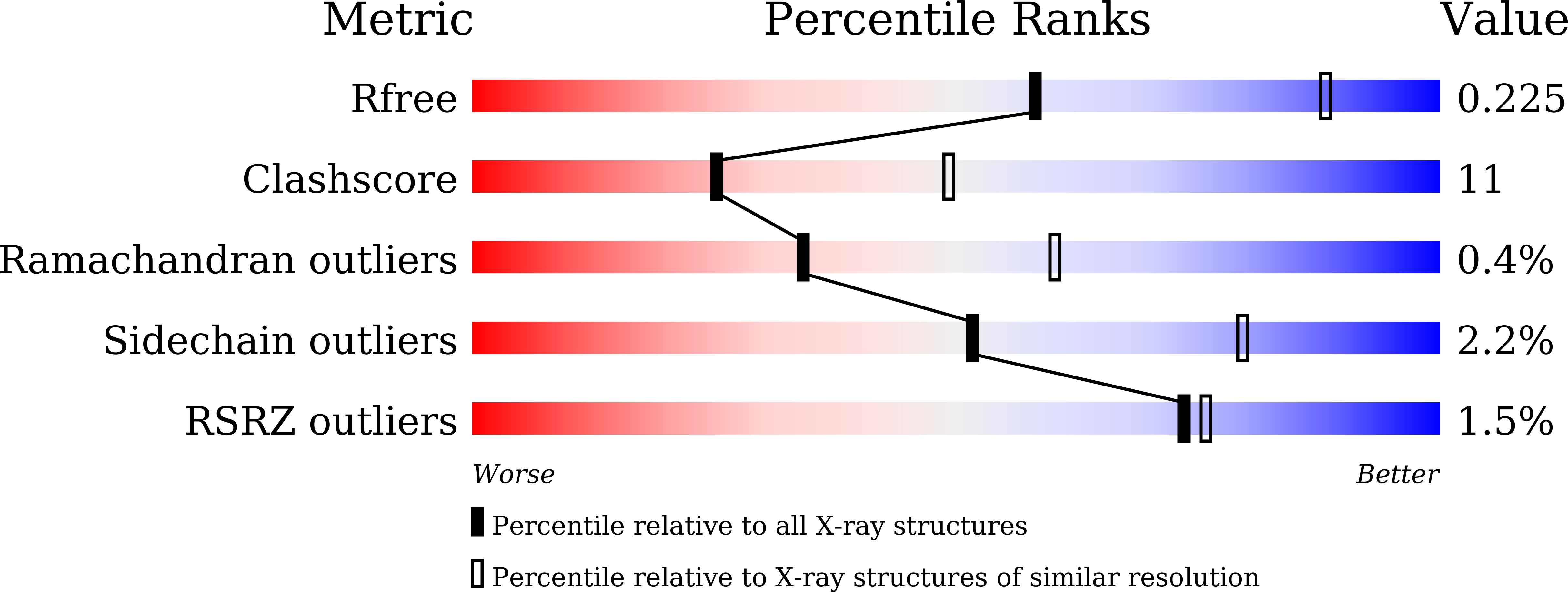
wwPDB Validation   3D Report Full Report


Ligand Structure Quality Assessment 


This is version 1.2 of the entry. See complete history


Literature

S19W, T27W, and N330Y mutations in ACE2 enhance SARS-CoV-2 S-RBD binding toward both wild-type and antibody-resistant viruses and its molecular basis.

Ye, F.Lin, X.Chen, Z.Yang, F.Lin, S.Yang, J.Chen, H.Sun, H.Wang, L.Wen, A.Zhang, X.Dai, Y.Cao, Y.Yang, J.Shen, G.Yang, L.Li, J.Wang, Z.Wang, W.Wei, X.Lu, G.

(2021) Signal Transduct Target Ther 6: 343-343

  • DOI: https://doi.org/10.1038/s41392-021-00756-4
  • Primary Citation of Related Structures:  
    7EFP, 7EFR

  • PubMed Abstract: 

    SARS-CoV-2 recognizes, via its spike receptor-binding domain (S-RBD), human angiotensin-converting enzyme 2 (ACE2) to initiate infection. Ecto-domain protein of ACE2 can therefore function as a decoy. Here we show that mutations of S19W, T27W, and N330Y in ACE2 could individually enhance SARS-CoV-2 S-RBD binding. Y330 could be synergistically combined with either W19 or W27, whereas W19 and W27 are mutually unbeneficial. The structures of SARS-CoV-2 S-RBD bound to the ACE2 mutants reveal that the enhanced binding is mainly contributed by the van der Waals interactions mediated by the aromatic side-chains from W19, W27, and Y330. While Y330 and W19/W27 are distantly located and devoid of any steric interference, W19 and W27 are shown to orient their side-chains toward each other and to cause steric conflicts, explaining their incompatibility. Finally, using pseudotyped SARS-CoV-2 viruses, we demonstrate that these residue substitutions are associated with dramatically improved entry-inhibition efficacy toward both wild-type and antibody-resistant viruses. Taken together, our biochemical and structural data have delineated the basis for the elevated S-RBD binding associated with S19W, T27W, and N330Y mutations in ACE2, paving the way for potential application of these mutants in clinical treatment of COVID-19.


  • Organizational Affiliation

    West China Hospital Emergency Department (WCHED), State Key Laboratory of Biotherapy, West China Hospital, Sichuan University, Chengdu, Sichuan, China.


Macromolecules
Find similar proteins by:  (by identity cutoff)  |  3D Structure
Entity ID: 1
MoleculeChains Sequence LengthOrganismDetailsImage
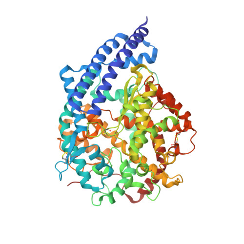
Processed angiotensin-converting enzyme 2599Homo sapiensMutation(s): 2 
Gene Names: ACE2UNQ868/PRO1885
EC: 3.4.17 (UniProt), 3.4.17.23 (UniProt)
UniProt & NIH Common Fund Data Resources
Find proteins for Q9BYF1 (Homo sapiens)
Explore Q9BYF1 
Go to UniProtKB:  Q9BYF1
PHAROS:  Q9BYF1
GTEx:  ENSG00000130234 
Entity Groups  
Sequence Clusters30% Identity50% Identity70% Identity90% Identity95% Identity100% Identity
UniProt GroupQ9BYF1
Glycosylation
Glycosylation Sites: 4Go to GlyGen: Q9BYF1-1
Sequence Annotations
Expand
  • Reference Sequence
Find similar proteins by:  (by identity cutoff)  |  3D Structure
Entity ID: 2
MoleculeChains Sequence LengthOrganismDetailsImage
Spike glycoprotein218Severe acute respiratory syndrome coronavirus 2Mutation(s): 0 
Gene Names: S2
UniProt
Find proteins for P0DTC2 (Severe acute respiratory syndrome coronavirus 2)
Explore P0DTC2 
Go to UniProtKB:  P0DTC2
Entity Groups  
Sequence Clusters30% Identity50% Identity70% Identity90% Identity95% Identity100% Identity
UniProt GroupP0DTC2
Glycosylation
Glycosylation Sites: 1Go to GlyGen: P0DTC2-1
Sequence Annotations
Expand
  • Reference Sequence
Experimental Data & Validation

Experimental Data

  • Method: X-RAY DIFFRACTION
  • Resolution: 2.70 Å
  • R-Value Free: 0.224 
  • R-Value Work: 0.184 
  • R-Value Observed: 0.186 
  • Space Group: P 43 21 2
Unit Cell:
Length ( Å )Angle ( ˚ )
a = 139.596α = 90
b = 139.596β = 90
c = 155.645γ = 90
Software Package:
Software NamePurpose
PHENIXrefinement
HKL-2000data scaling
PDB_EXTRACTdata extraction
HKL-2000data reduction
PHASERphasing
Cootmodel building

Structure Validation

View Full Validation Report


Ligand Structure Quality Assessment 


Entry History 

Deposition Data

Revision History  (Full details and data files)

  • Version 1.0: 2021-11-24
    Type: Initial release
  • Version 1.1: 2023-11-29
    Changes: Data collection, Refinement description
  • Version 1.2: 2024-10-23
    Changes: Structure summary