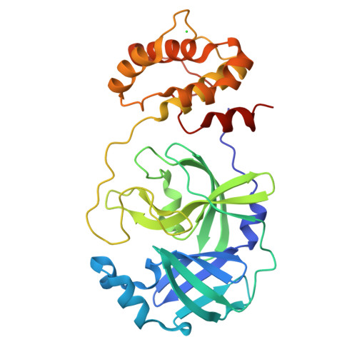
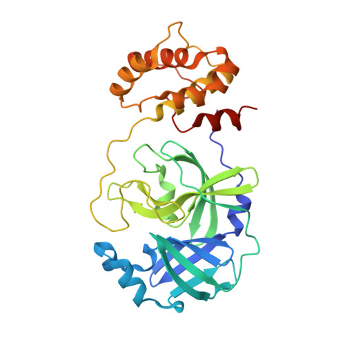
Detailed Insights into the Inhibitory Mechanism of New Ebselen Derivatives against Main Protease (M pro ) of Severe Acute Respiratory Syndrome Coronavirus-2 (SARS-CoV-2).
Sahoo, P., Lenka, D.R., Batabyal, M., Pain, P.K., Kumar, S., Manna, D., Kumar, A.(2023) Acs Pharmacol Transl Sci 6: 171-180
- PubMed: 36650888
- DOI: https://doi.org/10.1021/acsptsci.2c00203
- Primary Citation of Related Structures:
7W9G, 7XQ6, 7XQ7 - PubMed Abstract:
SARS-CoV-2 main protease (M pro /3CL pro ) is a crucial target for therapeutics, which is responsible for viral polyprotein cleavage and plays a vital role in virus replication and survival. Recent studies suggest that 2-phenylbenzisoselenazol-3(2 H )-one (ebselen) is a potent covalent inhibitor of M pro , which affects its enzymatic activity and virus survival. Herein, we synthesized various ebselen derivatives to understand the mechanism of M pro inhibition by ebselen. Using ebselen derivatives, we characterized the detailed interaction mechanism with M pro . We discovered that modification of the parent ebselen inhibitor with an electron-withdrawing group (NO 2 ) increases the inhibition efficacy by 2-fold. We also solved the structure of an M pro complex with an ebselen derivative showing the mechanism of inhibition by blocking the catalytic Cys145 of M pro . Using a combination of crystal structures and LC-MS data, we showed that M pro hydrolyzes the new ebselen derivative and leaves behind selenium (Se) bound with Cys145 of the catalytic dyad of M pro . We also described the binding profile of ebselen-based inhibitors using molecular modeling predictions supported by binding and inhibition assays. Furthermore, we have also solved the crystal structure of catalytically inactive mutant H41N-M pro , which represents the inactive state of the protein where the substrate binding pocket is blocked. The inhibited structure of H41N-M pro shows gatekeeper residues in the substrate binding pocket responsible for blocking the substrate binding; mutation of these gatekeeper residues leads to hyperactive M pro .
Organizational Affiliation:
Department of Biological Sciences, Indian Institute of Science Education and Research (IISER) Bhopal, Bhopal, Madhya Pradesh 462066, India.


