7Y9B

Crystal structure of the membrane (M) protein of a SARS-COV-2-related coronavirus


Experimental Data Snapshot

  • Method: X-RAY DIFFRACTION
  • Resolution: 3.21 Å
  • R-Value Free: 0.293 
  • R-Value Work: 0.271 
  • R-Value Observed: 0.272 

wwPDB Validation   3D Report Full Report


Ligand Structure Quality Assessment 


This is version 2.4 of the entry. See complete history


Literature

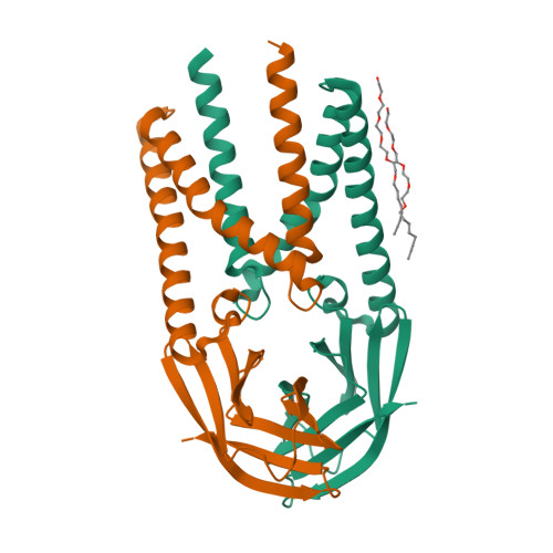
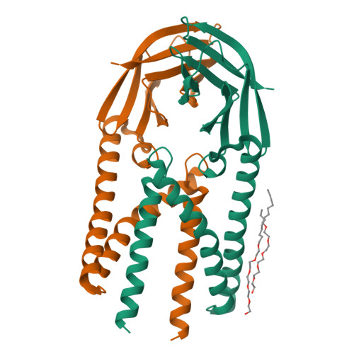
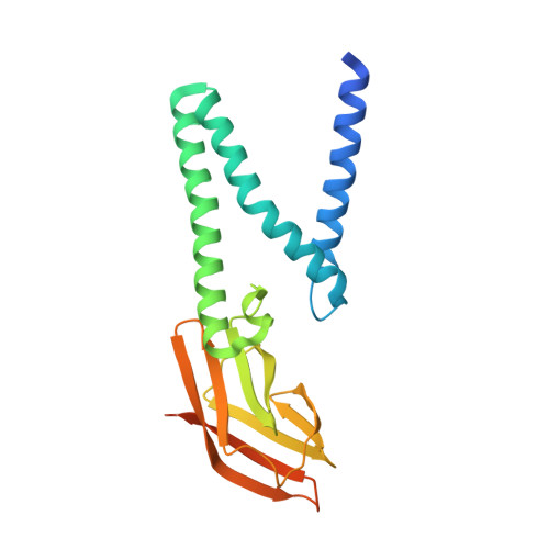
Crystal structure of the membrane (M) protein from a bat betacoronavirus.

Wang, X.Yang, Y.Sun, Z.Zhou, X.

(2023) PNAS Nexus 2: pgad021-pgad021

  • DOI: https://doi.org/10.1093/pnasnexus/pgad021
  • Primary Citation of Related Structures:  
    7Y96, 7Y9B

  • PubMed Abstract: 

    The membrane (M) protein is the most abundant structural protein of coronaviruses including MERS-CoV, SARS-CoV, and SARS-CoV-2, and plays a central role in virus assembly through its interaction with various partner proteins. However, mechanistic details about how M protein interacts with others remain elusive due to lack of high-resolution structures. Here, we present the first crystal structure of a betacoronavirus M protein from Pipistrellus bat coronavirus HKU5 (batCOV5-M), which is closely related to MERS-CoV, SARS-CoV, and SARS-CoV-2 M proteins. Furthermore, an interaction analysis indicates that the carboxy-terminus of the batCOV5 nucleocapsid (N) protein mediates its interaction with batCOV5-M. Combined with a computational docking analysis an M-N interaction model is proposed, providing insight into the mechanism of M protein-mediated protein interactions.


  • Organizational Affiliation

    State Key Laboratory of Biotherapy, Department of Integrated Traditional Chinese and Western Medicine, Rare Diseases Center, West China Hospital, Sichuan University, Chengdu, Sichuan 610041, China.


Macromolecules
Find similar proteins by:  (by identity cutoff)  |  3D Structure
Entity ID: 1
MoleculeChains Sequence LengthOrganismDetailsImage
Membrane protein
A, B, C, D
220Pipistrellus bat coronavirus HKU5Mutation(s): 0 
Gene Names: M5
Membrane Entity: Yes 
UniProt
Find proteins for A3EXD6 (Bat coronavirus HKU5)
Explore A3EXD6 
Go to UniProtKB:  A3EXD6
Entity Groups  
Sequence Clusters30% Identity50% Identity70% Identity90% Identity95% Identity100% Identity
UniProt GroupA3EXD6
Sequence Annotations
Expand
  • Reference Sequence
Experimental Data & Validation

Experimental Data

  • Method: X-RAY DIFFRACTION
  • Resolution: 3.21 Å
  • R-Value Free: 0.293 
  • R-Value Work: 0.271 
  • R-Value Observed: 0.272 
  • Space Group: P 1 21 1
Unit Cell:
Length ( Å )Angle ( ˚ )
a = 76.569α = 90
b = 66.571β = 109.83
c = 112.628γ = 90
Software Package:
Software NamePurpose
PHENIXrefinement
XDSdata reduction
Aimlessdata scaling
PHASERphasing
PDB_EXTRACTdata extraction

Structure Validation

View Full Validation Report


Ligand Structure Quality Assessment 


Entry History & Funding Information

Deposition Data


Funding OrganizationLocationGrant Number
National Natural Science Foundation of China (NSFC)China31770783

Revision History  (Full details and data files)

  • Version 1.0: 2022-08-17
    Type: Initial release
  • Version 2.0: 2022-11-23
    Type: Coordinate replacement
    Reason: Ligand identity
    Changes: Atomic model, Data collection, Database references, Derived calculations, Non-polymer description, Refinement description, Source and taxonomy, Structure summary
  • Version 2.1: 2023-03-01
    Changes: Database references
  • Version 2.2: 2023-03-22
    Changes: Database references
  • Version 2.3: 2023-05-31
    Changes: Database references
  • Version 2.4: 2023-11-29
    Changes: Data collection, Refinement description