8GZ5

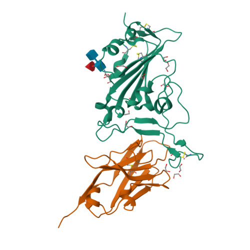
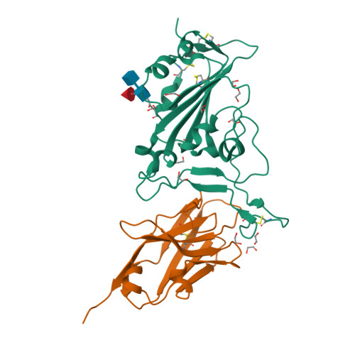
Crystal structure of neutralizing VHH P17 in complex with SARS-CoV-2 Alpha variant spike receptor-binding domain


Experimental Data Snapshot

  • Method: X-RAY DIFFRACTION
  • Resolution: 1.70 Å
  • R-Value Free: 0.189 
  • R-Value Work: 0.171 
  • R-Value Observed: 0.172 

Starting Models: experimental
View more details

wwPDB Validation   3D Report Full Report


This is version 1.3 of the entry. See complete history


Literature

Structural insights into the rational design of a nanobody that binds with high affinity to the SARS-CoV-2 spike variant.

Yamaguchi, K.Anzai, I.Maeda, R.Moriguchi, M.Watanabe, T.Imura, A.Takaori-Kondo, A.Inoue, T.

(2023) J Biochem 173: 115-127

  • DOI: https://doi.org/10.1093/jb/mvac096
  • Primary Citation of Related Structures:  
    8GZ5, 8GZ6

  • PubMed Abstract: 

    The continuous emergence of severe acute respiratory syndrome coronavirus-2 (SARS-CoV-2) variants associated with the adaptive evolution of the virus is prolonging the global coronavirus disease 2019 (COVID-19) pandemic. The modification of neutralizing antibodies based on structural information is expected to be a useful approach to rapidly combat emerging variants. A dimerized variable domain of heavy chain of heavy chain antibody (VHH) P17 that has highly potent neutralizing activity against SARS-CoV-2 has been reported but the mode of interaction with the epitope remains unclear. Here, we report the X-ray crystal structure of the complex of monomerized P17 bound to the SARS-CoV-2 receptor binding domain (RBD) and investigated the binding activity of P17 toward various variants of concern (VOCs) using kinetics measurements. The structure revealed details of the binding interface and showed that P17 had an appropriate linker length to have an avidity effect and recognize a wide range of RBD orientations. Furthermore, we identified mutations in known VOCs that decrease the binding affinity of P17 and proposed methods for the acquisition of affinity toward the Omicron RBD because Omicron is currently the most predominant VOC. This study provides information for the rational design of effective VHHs for emerging VOCs.


  • Organizational Affiliation

    Graduate School of Pharmaceutical Sciences, Osaka University, 1-6 Yamada-oka, Suita, Osaka 565-0871, Japan.


Macromolecules
Find similar proteins by:  (by identity cutoff)  |  3D Structure
Entity ID: 1
MoleculeChains Sequence LengthOrganismDetailsImage
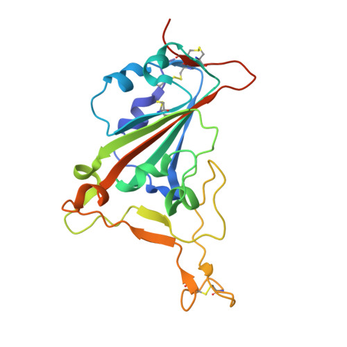
Spike protein S1206Severe acute respiratory syndrome coronavirus 2Mutation(s): 1 
Gene Names: S2
UniProt
Find proteins for P0DTC2 (Severe acute respiratory syndrome coronavirus 2)
Explore P0DTC2 
Go to UniProtKB:  P0DTC2
Entity Groups  
Sequence Clusters30% Identity50% Identity70% Identity90% Identity95% Identity100% Identity
UniProt GroupP0DTC2
Glycosylation
Glycosylation Sites: 1Go to GlyGen: P0DTC2-1
Sequence Annotations
Expand
  • Reference Sequence
Find similar proteins by:  (by identity cutoff)  |  3D Structure
Entity ID: 2
MoleculeChains Sequence LengthOrganismDetailsImage
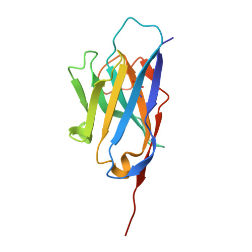
Nanobody P17126Vicugna pacosMutation(s): 0 
Entity Groups  
Sequence Clusters30% Identity50% Identity70% Identity90% Identity95% Identity100% Identity
Sequence Annotations
Expand
  • Reference Sequence
Oligosaccharides

Help

Entity ID: 3
MoleculeChains Length2D Diagram Glycosylation3D Interactions
2-acetamido-2-deoxy-beta-D-glucopyranose-(1-4)-[alpha-L-fucopyranose-(1-6)]2-acetamido-2-deoxy-beta-D-glucopyranose
C
3N-Glycosylation
Glycosylation Resources
GlyTouCan:  G21290RB
GlyCosmos:  G21290RB
GlyGen:  G21290RB
Small Molecules
Ligands 1 Unique
IDChains Name / Formula / InChI Key2D Diagram3D Interactions
EDO
Query on EDO

Download Ideal Coordinates CCD File 
D [auth A]
E [auth A]
F [auth A]
G [auth A]
H [auth A]
D [auth A],
E [auth A],
F [auth A],
G [auth A],
H [auth A],
I [auth A],
J [auth A],
K [auth A],
L [auth A],
M [auth A],
N [auth B],
O [auth B]
1,2-ETHANEDIOL
C2 H6 O2
LYCAIKOWRPUZTN-UHFFFAOYSA-N
Experimental Data & Validation

Experimental Data

  • Method: X-RAY DIFFRACTION
  • Resolution: 1.70 Å
  • R-Value Free: 0.189 
  • R-Value Work: 0.171 
  • R-Value Observed: 0.172 
  • Space Group: P 32 2 1
Unit Cell:
Length ( Å )Angle ( ˚ )
a = 82.68α = 90
b = 82.68β = 90
c = 162.479γ = 120
Software Package:
Software NamePurpose
PHENIXrefinement
XDSdata reduction
Aimlessdata scaling
PHASERphasing

Structure Validation

View Full Validation Report


Entry History & Funding Information

Deposition Data


Funding OrganizationLocationGrant Number
Japan Science and TechnologyJapanJPMJOP1861
Japan Agency for Medical Research and Development (AMED)JapanJP20fk018517
Other governmentJapanKYOTO Industrial Support Organization 21

Revision History  (Full details and data files)

  • Version 1.0: 2022-12-07
    Type: Initial release
  • Version 1.1: 2023-02-15
    Changes: Database references
  • Version 1.2: 2023-11-29
    Changes: Data collection, Refinement description
  • Version 1.3: 2024-10-23
    Changes: Structure summary