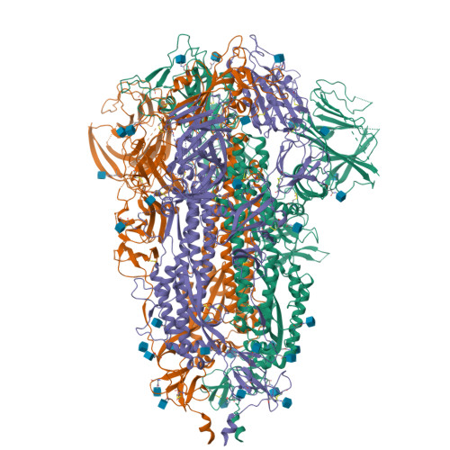
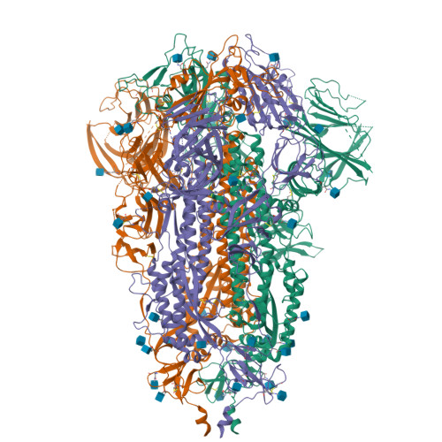
8PSD

SARS-CoV-2 XBB 1.0 closed conformation.


Experimental Data Snapshot

  • Method: ELECTRON MICROSCOPY
  • Resolution: 2.90 Å
  • Aggregation State: PARTICLE 
  • Reconstruction Method: SINGLE PARTICLE 

Starting Model: in silico
View more details

wwPDB Validation   3D Report Full Report


This is version 1.2 of the entry. See complete history


Literature

Broadly potent anti-SARS-CoV-2 antibody shares 93% of epitope with ACE2 and provides full protection in monkeys.

Fenwick, C.Turelli, P.Duhoo, Y.Lau, K.Herate, C.Marlin, R.Lamrayah, M.Campos, J.Esteves-Leuenberger, L.Farina, A.Raclot, C.Genet, V.Fiscalini, F.Cesborn, J.Perez, L.Dereuddre-Bosquet, N.Contreras, V.Lheureux, K.Relouzat, F.Abdelnabi, R.Leyssen, P.Levy, Y.Pojer, F.Le Grand, R.Trono, D.Pantaleo, G.

(2023) J Infect 87: 524-537

  • DOI: https://doi.org/10.1016/j.jinf.2023.10.008
  • Primary Citation of Related Structures:  
    8PQ2, 8PSD

  • PubMed Abstract: 

    Due to the rapid evolution of SARS-CoV-2 to variants with reduced sensitivity to vaccine-induced humoral immunity and the near complete loss of protective efficacy of licensed therapeutic monoclonal antibodies, we isolated a potent, broad-spectrum neutralizing antibody that could potentially provide prophylactic protection to immunocompromised patient populations. Spike-specific B-cell clones isolated from a vaccinated post-infected donor were profiled for those producing potent neutralizing antibodies against a panel of SARS-CoV-2 variants. The P4J15 antibody was further characterized to define the structural binding epitope, viral resistance, and in vivo efficacy. The P4J15 mAb shows <20 ng/ml neutralizing activity against all variants including the latest XBB.2.3 and EG.5.1 sub-lineages. Structural studies of P4J15 in complex with Omicron XBB.1 Spike show that the P4J15 epitope shares ∼93% of its buried surface area with the ACE2 contact region, consistent with an ACE2 mimetic antibody. In vitro selection of SARS-CoV-2 mutants escaping P4J15 neutralization showed reduced infectivity, poor ACE2 binding, and mutations are rare in public sequence databases. Using a SARS-CoV-2 XBB.1.5 monkey challenge model, P4J15-LS confers complete prophylactic protection with an exceptionally long in vivo half-life of 43 days. The P4J15 mAb has potential as a broad-spectrum anti-SARS-CoV-2 drug for prophylactic protection of at-risk patient populations.


  • Organizational Affiliation

    Service of Immunology and Allergy, Department of Medicine, Lausanne University Hospital and University of Lausanne, Lausanne, Switzerland.


Macromolecules
Find similar proteins by:  (by identity cutoff)  |  3D Structure
Entity ID: 1
MoleculeChains Sequence LengthOrganismDetailsImage
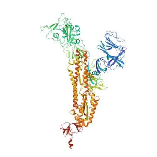
Spike glycoproteinA [auth C],
B [auth A],
C [auth B]
1,204Severe acute respiratory syndrome coronavirus 2Mutation(s): 0 
Gene Names: S2
UniProt
Find proteins for P0DTC2 (Severe acute respiratory syndrome coronavirus 2)
Explore P0DTC2 
Go to UniProtKB:  P0DTC2
Entity Groups  
Sequence Clusters30% Identity50% Identity70% Identity90% Identity95% Identity100% Identity
UniProt GroupP0DTC2
Glycosylation
Glycosylation Sites: 10Go to GlyGen: P0DTC2-1
Sequence Annotations
Expand
  • Reference Sequence
Small Molecules
Ligands 1 Unique
IDChains Name / Formula / InChI Key2D Diagram3D Interactions
NAG
Query on NAG

Download Ideal Coordinates CCD File 
AA [auth A]
BA [auth B]
CA [auth B]
D [auth C]
DA [auth B]
AA [auth A],
BA [auth B],
CA [auth B],
D [auth C],
DA [auth B],
E [auth C],
EA [auth B],
F [auth C],
FA [auth B],
G [auth C],
GA [auth B],
H [auth C],
HA [auth B],
I [auth C],
IA [auth B],
J [auth C],
JA [auth B],
K [auth C],
KA [auth B],
L [auth C],
LA [auth B],
M [auth C],
MA [auth B],
N [auth C],
O [auth C],
P [auth C],
Q [auth A],
R [auth A],
S [auth A],
T [auth A],
U [auth A],
V [auth A],
W [auth A],
X [auth A],
Y [auth A],
Z [auth A]
2-acetamido-2-deoxy-beta-D-glucopyranose
C8 H15 N O6
OVRNDRQMDRJTHS-FMDGEEDCSA-N
Experimental Data & Validation

Experimental Data

  • Method: ELECTRON MICROSCOPY
  • Resolution: 2.90 Å
  • Aggregation State: PARTICLE 
  • Reconstruction Method: SINGLE PARTICLE 
EM Software:
TaskSoftware PackageVersion
MODEL REFINEMENTPHENIX
RECONSTRUCTIONcryoSPARC4.1

Structure Validation

View Full Validation Report


Entry History & Funding Information

Deposition Data


Funding OrganizationLocationGrant Number
Swiss National Science FoundationSwitzerland--

Revision History  (Full details and data files)

  • Version 1.0: 2023-11-01
    Type: Initial release
  • Version 1.1: 2023-12-06
    Changes: Database references
  • Version 1.2: 2024-11-13
    Changes: Data collection, Structure summary