8PVM

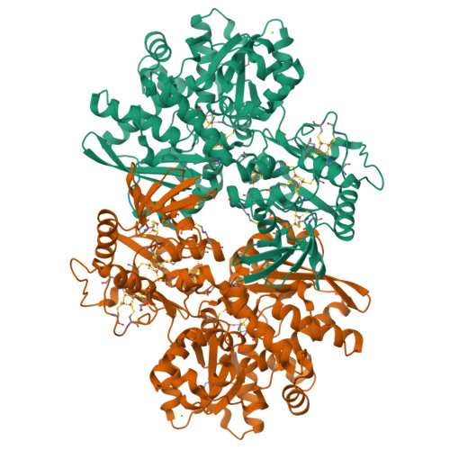
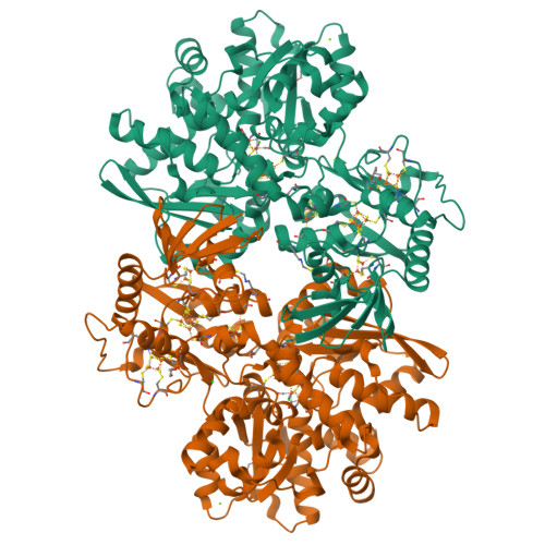
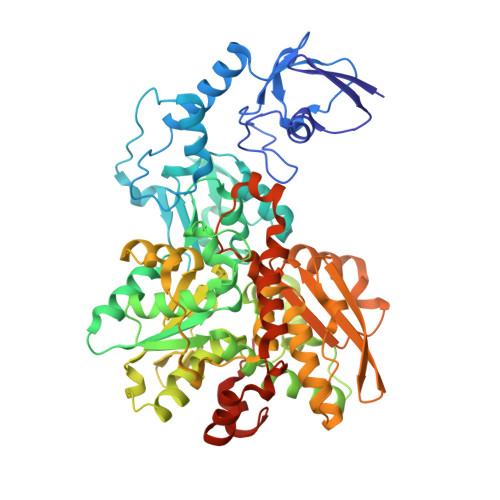
formaldehyde-inhibited [FeFe]-hydrogenase CpI from Clostridium pasteurianum, variant C299D


Experimental Data Snapshot

  • Method: X-RAY DIFFRACTION
  • Resolution: 1.38 Å
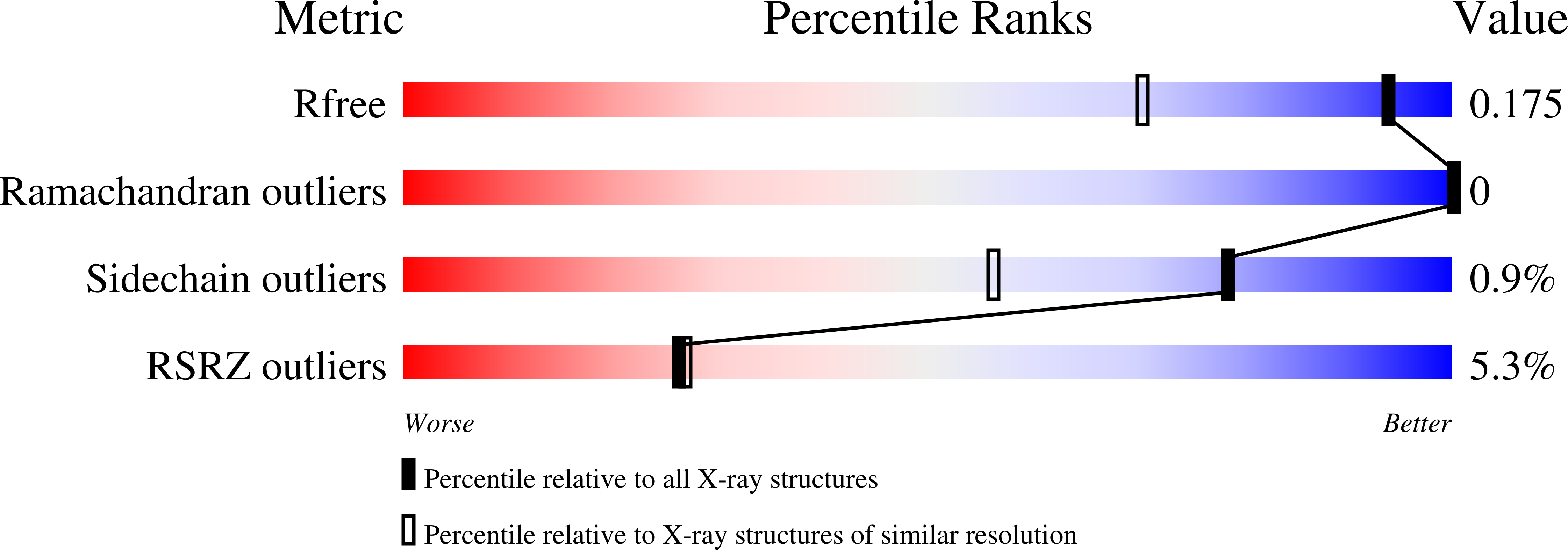
  • R-Value Free: 0.175 
  • R-Value Work: 0.143 
  • R-Value Observed: 0.144 

Starting Model: experimental
View more details

wwPDB Validation   3D Report Full Report


Ligand Structure Quality Assessment 


This is version 1.3 of the entry. See complete history


Literature

Insights into the Molecular Mechanism of Formaldehyde Inhibition of [FeFe]-Hydrogenases.

Duan, J.Veliju, A.Lampret, O.Liu, L.Yadav, S.Apfel, U.P.Armstrong, F.A.Hemschemeier, A.Hofmann, E.

(2023) J Am Chem Soc 145: 26068-26074

  • DOI: https://doi.org/10.1021/jacs.3c07800
  • Primary Citation of Related Structures:  
    8PVM, 8QM3

  • PubMed Abstract: 

    [FeFe]-hydrogenases are efficient H 2 converting biocatalysts that are inhibited by formaldehyde (HCHO). The molecular mechanism of this inhibition has so far not been experimentally solved. Here, we obtained high-resolution crystal structures of the HCHO-treated [FeFe]-hydrogenase CpI from Clostridium pasteurianum , showing HCHO reacts with the secondary amine base of the catalytic cofactor and the cysteine C299 of the proton transfer pathway which both are very important for catalytic turnover. Kinetic assays via protein film electrochemistry show the CpI variant C299D is significantly less inhibited by HCHO, corroborating the structural results. By combining our data from protein crystallography, site-directed mutagenesis and protein film electrochemistry, a reaction mechanism involving the cofactor's amine base, the thiol group of C299 and HCHO can be deduced. In addition to the specific case of [FeFe]-hydrogenases, our study provides additional insights into the reactions between HCHO and protein molecules.


  • Organizational Affiliation

    Photobiotechnology, Faculty of Biology and Biotechnology, Ruhr University Bochum, 44801 Bochum, Germany.


Macromolecules
Find similar proteins by:  (by identity cutoff)  |  3D Structure
Entity ID: 1
MoleculeChains Sequence LengthOrganismDetailsImage
Iron hydrogenase 1
A, B
584Clostridium pasteurianumMutation(s): 0 
EC: 1.12.7.2
UniProt
Find proteins for P29166 (Clostridium pasteurianum)
Explore P29166 
Go to UniProtKB:  P29166
Entity Groups  
Sequence Clusters30% Identity50% Identity70% Identity90% Identity95% Identity100% Identity
UniProt GroupP29166
Sequence Annotations
Expand
  • Reference Sequence
Small Molecules
Ligands 8 Unique
IDChains Name / Formula / InChI Key2D Diagram3D Interactions
402 (Subject of Investigation/LOI)
Query on 402

Download Ideal Coordinates CCD File 
C [auth A],
R [auth B]
dicarbonyl[bis(cyanide-kappaC)]-mu-(iminodimethanethiolatato-1kappaS:2kappaS)-mu-(oxomethylidene)diiron(2+)
C7 H5 Fe2 N3 O3 S2
LJPDYWPSPOWMIB-UHFFFAOYSA-N
SF4
Query on SF4

Download Ideal Coordinates CCD File 
D [auth A]
E [auth A]
F [auth A]
G [auth A]
S [auth B]
D [auth A],
E [auth A],
F [auth A],
G [auth A],
S [auth B],
T [auth B],
U [auth B],
V [auth B]
IRON/SULFUR CLUSTER
Fe4 S4
LJBDFODJNLIPKO-UHFFFAOYSA-N
FES
Query on FES

Download Ideal Coordinates CCD File 
H [auth A],
W [auth B]
FE2/S2 (INORGANIC) CLUSTER
Fe2 S2
NIXDOXVAJZFRNF-UHFFFAOYSA-N
GOL
Query on GOL

Download Ideal Coordinates CCD File 
AA [auth B],
J [auth A],
K [auth A],
Z [auth B]
GLYCEROL
C3 H8 O3
PEDCQBHIVMGVHV-UHFFFAOYSA-N
CL
Query on CL

Download Ideal Coordinates CCD File 
FA [auth B]
GA [auth B]
HA [auth B]
O [auth A]
P [auth A]
FA [auth B],
GA [auth B],
HA [auth B],
O [auth A],
P [auth A],
Q [auth A]
CHLORIDE ION
Cl
VEXZGXHMUGYJMC-UHFFFAOYSA-M
FOR
Query on FOR

Download Ideal Coordinates CCD File 
BA [auth B],
L [auth A]
FORMYL GROUP
C H2 O
WSFSSNUMVMOOMR-UHFFFAOYSA-N
MG
Query on MG

Download Ideal Coordinates CCD File 
CA [auth B],
DA [auth B],
EA [auth B],
M [auth A],
N [auth A]
MAGNESIUM ION
Mg
JLVVSXFLKOJNIY-UHFFFAOYSA-N
74C
Query on 74C

Download Ideal Coordinates CCD File 
I [auth A],
X [auth B],
Y [auth B]
methyl radical
C H3
VNWKTOKETHGBQD-UHFFFAOYSA-N
Experimental Data & Validation

Experimental Data

  • Method: X-RAY DIFFRACTION
  • Resolution: 1.38 Å
  • R-Value Free: 0.175 
  • R-Value Work: 0.143 
  • R-Value Observed: 0.144 
  • Space Group: P 1 21 1
Unit Cell:
Length ( Å )Angle ( ˚ )
a = 87.65α = 90
b = 69.83β = 102.74
c = 102.89γ = 90
Software Package:
Software NamePurpose
PHENIXrefinement
XSCALEdata scaling
XDSdata reduction
PHENIXphasing

Structure Validation

View Full Validation Report


Ligand Structure Quality Assessment 


Entry History & Funding Information

Deposition Data


Funding OrganizationLocationGrant Number
German Research Foundation (DFG)Germany--

Revision History  (Full details and data files)

  • Version 1.0: 2023-11-29
    Type: Initial release
  • Version 1.1: 2023-12-06
    Changes: Database references
  • Version 1.2: 2023-12-13
    Changes: Database references
  • Version 1.3: 2024-10-23
    Changes: Structure summary