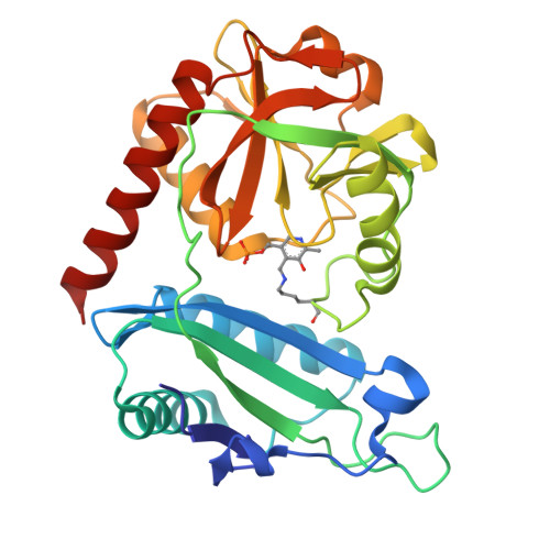
8RAF

Crystal structure of D-amino acid transaminase from Haliscomenobacter hydrossis point mutant R90I (holo form)


Experimental Data Snapshot

  • Method: X-RAY DIFFRACTION
  • Resolution: 2.00 Å
  • R-Value Free: 0.236 
  • R-Value Work: 0.194 
  • R-Value Observed: 0.196 

Starting Model: experimental
View more details

wwPDB Validation   3D Report Full Report


Ligand Structure Quality Assessment 


This is version 1.2 of the entry. See complete history


Literature

Multifunctionality of arginine residues in the active sites of non-canonical d-amino acid transaminases.

Bakunova, A.K.Matyuta, I.O.Minyaev, M.E.Isaikina, T.Y.Boyko, K.M.Popov, V.O.Bezsudnova, E.Y.

(2024) Arch Biochem Biophys 756: 110011-110011

  • DOI: https://doi.org/10.1016/j.abb.2024.110011
  • Primary Citation of Related Structures:  
    8RAF, 8RAI

  • PubMed Abstract: 

    Structure-function relationships are key to understanding enzyme mechanisms, controlling enzyme activities, and designing biocatalysts. Here, we investigate the functions of arginine residues in the active sites of pyridoxal-5'-phosphate (PLP)-dependent non-canonical d-amino acid transaminases, focusing on the analysis of a transaminase from Haliscomenobacter hydrossis. Our results show that the tandem of arginine residues R28* and R90, which form the conserved R-[RK] motif in non-canonical d-amino acid transaminases, not only facilitates effective substrate binding but also regulates the catalytic properties of PLP. Non-covalent interactions between residues R28*, R90, and Y147 strengthen the hydrogen bond between Y147 and PLP, thereby maintaining the reactivity of the cofactor. Next, the R90 residue contributes to the stability of the holoenzyme. Finally, the R90I substitution induces structural changes that lead to substrate promiscuity, as evidenced by the effective binding of substrates with and without the α-carboxylate group. This study sheds light on the structural determinants of the activity of non-canonical d-amino acid transaminases. Understanding the structural basis of the active site plasticity in the non-canonical transaminase from H. hydrossis, which is characterized by effective conversion of d-amino acids and α-keto acids, may help to tailor it for industrial applications.


  • Organizational Affiliation

    Bach Institute of Biochemistry, Research Centre of Biotechnology of the Russian Academy of Sciences, Leninsky Ave. 33, bld. 2, 119071, Moscow, Russia. Electronic address: a.bakunova@fbras.ru.


Macromolecules
Find similar proteins by:  (by identity cutoff)  |  3D Structure
Entity ID: 1
MoleculeChains Sequence LengthOrganismDetailsImage
Aminotransferase class IV283Haliscomenobacter hydrossis DSM 1100Mutation(s): 1 
Gene Names: Halhy_2446
UniProt
Find proteins for F4KWH0 (Haliscomenobacter hydrossis (strain ATCC 27775 / DSM 1100 / LMG 10767 / O))
Explore F4KWH0 
Go to UniProtKB:  F4KWH0
Entity Groups  
Sequence Clusters30% Identity50% Identity70% Identity90% Identity95% Identity100% Identity
UniProt GroupF4KWH0
Sequence Annotations
Expand
  • Reference Sequence
Small Molecules
Ligands 1 Unique
IDChains Name / Formula / InChI Key2D Diagram3D Interactions
PLP (Subject of Investigation/LOI)
Query on PLP

Download Ideal Coordinates CCD File 
B [auth A]PYRIDOXAL-5'-PHOSPHATE
C8 H10 N O6 P
NGVDGCNFYWLIFO-UHFFFAOYSA-N
Experimental Data & Validation

Experimental Data

  • Method: X-RAY DIFFRACTION
  • Resolution: 2.00 Å
  • R-Value Free: 0.236 
  • R-Value Work: 0.194 
  • R-Value Observed: 0.196 
  • Space Group: C 1 2 1
Unit Cell:
Length ( Å )Angle ( ˚ )
a = 85.392α = 90
b = 72.627β = 100.8
c = 52.244γ = 90
Software Package:
Software NamePurpose
REFMACrefinement
Aimlessdata scaling
CrysalisProdata reduction
REFMACphasing
PDB_EXTRACTdata extraction

Structure Validation

View Full Validation Report


Ligand Structure Quality Assessment 


Entry History & Funding Information

Deposition Data


Funding OrganizationLocationGrant Number
Russian Science FoundationRussian Federation23-74-30004

Revision History  (Full details and data files)

  • Version 1.0: 2023-12-27
    Type: Initial release
  • Version 1.1: 2024-05-01
    Changes: Database references
  • Version 1.2: 2024-05-08
    Changes: Database references