1E2J

The nucleoside binding site of Herpes simplex type 1 thymidine kinase analyzed by X-ray crystallography


Experimental Data Snapshot

  • Method: X-RAY DIFFRACTION
  • Resolution: 2.50 Å
  • R-Value Free: 0.273 
  • R-Value Work: 0.212 

Starting Model: experimental
View more details

wwPDB Validation   3D Report Full Report


Ligand Structure Quality Assessment 


This is version 1.4 of the entry. See complete history


Literature

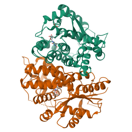
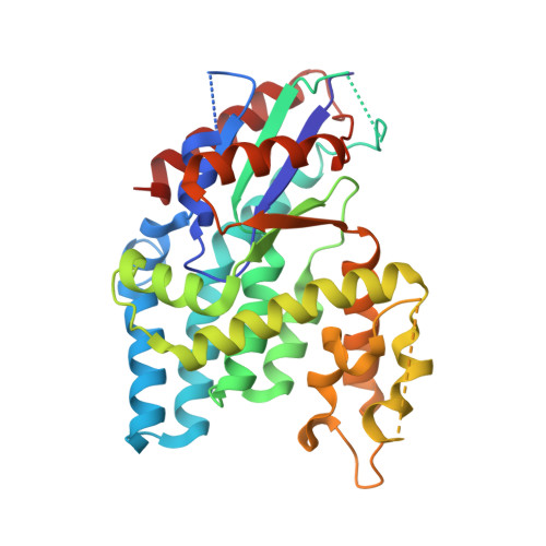
Nucleoside Binding Site of Herpes Simplex Type 1 Thymidine Kinase Analyzed by X-Ray Crystallography

Vogt, J.Perozzo, R.Pautsch, A.Prota, A.Schelling, P.Pilger, B.Folkers, G.Scapozza, L.Schulz, G.E.

(2000) Proteins 41: 545

  • DOI: https://doi.org/10.1002/1097-0134(20001201)41:4<545::aid-prot110>3.0.co;2-8
  • Primary Citation of Related Structures:  
    1E2H, 1E2I, 1E2J

  • PubMed Abstract: 

    The crystal structures of the full-length Herpes simplex virus type 1 thymidine kinase in its unligated form and in a complex with an adenine analogue have been determined at 1.9 A resolution. The unligated enzyme contains four water molecules in the thymidine pocket and reveals a small induced fit on substrate binding. The structure of the ligated enzyme shows for the first time a bound adenine analogue after numerous complexes with thymine and guanine analogues have been reported. The adenine analogue constitutes a new lead compound for enzyme-prodrug gene therapy. In addition, the structure of mutant Q125N modifying the binding site of the natural substrate thymidine in complex with this substrate has been established at 2.5 A resolution. It reveals that neither the binding mode of thymidine nor the polypeptide backbone conformation is altered, except that the two major hydrogen bonds to thymidine are replaced by a single water-mediated hydrogen bond, which improves the relative acceptance of the prodrugs aciclovir and ganciclovir compared with the natural substrate. Accordingly, the mutant structure represents a first step toward improving the virus-directed enzyme-prodrug gene therapy by enzyme engineering.


  • Organizational Affiliation

    Institut für Organische Chemie und Biochemie, Albert-Ludwigs-Universität, Freiburg im Breisgau, Germany.


Macromolecules
Find similar proteins by:  (by identity cutoff)  |  3D Structure
Entity ID: 1
MoleculeChains Sequence LengthOrganismDetailsImage
THYMIDINE KINASE
A, B
331Human alphaherpesvirus 1 strain 17Mutation(s): 1 
EC: 2.7.1.21
UniProt
Find proteins for P0DTH5 (Human herpesvirus 1 (strain 17))
Explore P0DTH5 
Go to UniProtKB:  P0DTH5
Entity Groups  
Sequence Clusters30% Identity50% Identity70% Identity90% Identity95% Identity100% Identity
UniProt GroupP0DTH5
Sequence Annotations
Expand
  • Reference Sequence
Small Molecules
Binding Affinity Annotations 
IDSourceBinding Affinity
THM BindingDB:  1E2J IC50: 1000 (nM) from 1 assay(s)
-TΔS: min: 27.18, max: 50.09 (kJ/mol) from 4 assay(s)
ΔH: min: -7.99e+1, max: -5.71e+1 (kJ/mol) from 4 assay(s)
ΔG: min: -3.02e+1, max: -2.94e+1 (kJ/mol) from 3 assay(s)
Experimental Data & Validation

Experimental Data

  • Method: X-RAY DIFFRACTION
  • Resolution: 2.50 Å
  • R-Value Free: 0.273 
  • R-Value Work: 0.212 
  • Space Group: C 2 2 21
Unit Cell:
Length ( Å )Angle ( ˚ )
a = 113.4α = 90
b = 117.5β = 90
c = 108.6γ = 90
Software Package:
Software NamePurpose
REFMACrefinement
XDSdata reduction
SCALAdata scaling
AMoREphasing

Structure Validation

View Full Validation Report


Ligand Structure Quality Assessment 


Entry History 

Deposition Data

Revision History  (Full details and data files)

  • Version 1.0: 2000-11-06
    Type: Initial release
  • Version 1.1: 2011-05-08
    Changes: Version format compliance
  • Version 1.2: 2011-07-13
    Changes: Version format compliance
  • Version 1.3: 2017-07-05
    Changes: Data collection
  • Version 1.4: 2023-12-06
    Changes: Data collection, Database references, Derived calculations, Other, Refinement description