1MOF

COAT PROTEIN


Experimental Data Snapshot

  • Method: X-RAY DIFFRACTION
  • Resolution: 1.70 Å
  • R-Value Free: 0.234 
  • R-Value Work: 0.169 
  • R-Value Observed: 0.169 

wwPDB Validation   3D Report Full Report


This is version 1.5 of the entry. See complete history


Literature

Retrovirus envelope domain at 1.7 angstrom resolution.

Fass, D.Harrison, S.C.Kim, P.S.

(1996) Nat Struct Biol 3: 465-469

  • DOI: https://doi.org/10.1038/nsb0596-465
  • Primary Citation of Related Structures:  
    1MOF

  • PubMed Abstract: 

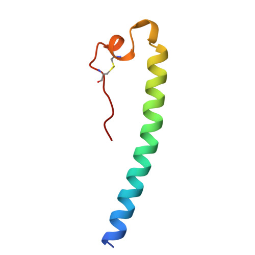
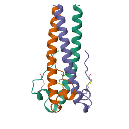
    We report the crystal structure of an extraviral segment of a retrovirus envelope protein, the Moloney murine leukemia virus (MoMuLV) transmembrane (TM) subunit. This segment, which comprises a region of the MoMuLV TM protein analogous to that contained within the X-ray crystal structure of low-pH converted influenza hemagglutinin, contains a trimeric coiled coil, with a hydrophobic cluster at its base and a strand that packs in an antiparallel orientation against the coiled coil. This structure gives the first high-resolution insight into the retrovirus surface and serves as a model for a wide range of viral fusion proteins; key residues in this structure are conserved among C- and D-type retroviruses and the filovirus ebola.


  • Organizational Affiliation

    Howard Hughes Medical Institute, Cambridge, Massachusetts, 02142, USA.


Macromolecules
Find similar proteins by:  (by identity cutoff)  |  3D Structure
Entity ID: 1
MoleculeChains Sequence LengthOrganismDetailsImage
MOLONEY MURINE LEUKEMIA VIRUS P1555Moloney murine leukemia virusMutation(s): 1 
UniProt
Find proteins for P03385 (Moloney murine leukemia virus (isolate Shinnick))
Explore P03385 
Go to UniProtKB:  P03385
Entity Groups  
Sequence Clusters30% Identity50% Identity70% Identity90% Identity95% Identity100% Identity
UniProt GroupP03385
Sequence Annotations
Expand
  • Reference Sequence
Small Molecules
Ligands 1 Unique
IDChains Name / Formula / InChI Key2D Diagram3D Interactions
CL
Query on CL

Download Ideal Coordinates CCD File 
B [auth A]CHLORIDE ION
Cl
VEXZGXHMUGYJMC-UHFFFAOYSA-M
Experimental Data & Validation

Experimental Data

  • Method: X-RAY DIFFRACTION
  • Resolution: 1.70 Å
  • R-Value Free: 0.234 
  • R-Value Work: 0.169 
  • R-Value Observed: 0.169 
  • Space Group: P 21 3
Unit Cell:
Length ( Å )Angle ( ˚ )
a = 53.1α = 90
b = 53.1β = 90
c = 53.1γ = 90
Software Package:
Software NamePurpose
BUDDHAdata collection
X-PLORrefinement
BUDDHAdata reduction

Structure Validation

View Full Validation Report


Entry History 

Deposition Data

Revision History  (Full details and data files)

  • Version 1.0: 1996-10-14
    Type: Initial release
  • Version 1.1: 2008-03-24
    Changes: Version format compliance
  • Version 1.2: 2011-07-13
    Changes: Derived calculations, Version format compliance
  • Version 1.3: 2018-06-27
    Changes: Data collection, Database references, Other
  • Version 1.4: 2022-12-21
    Changes: Database references, Derived calculations
  • Version 1.5: 2024-10-30
    Changes: Data collection, Structure summary