1O7P | pdb_00001o7p

NAPHTHALENE 1,2-DIOXYGENASE, PRODUCT COMPLEX


Experimental Data Snapshot

  • Method: X-RAY DIFFRACTION
  • Resolution: 1.95 Å
  • R-Value Free: 
    0.219 (Depositor), 0.210 (DCC) 
  • R-Value Work: 
    0.184 (Depositor), 0.190 (DCC) 

Starting Model: experimental
View more details

wwPDB Validation   3D Report Full Report


Ligand Structure Quality Assessment 

Created with Raphaël 2.3.0Worse 01 BetterLigand structure goodness of fit to experimental dataBest fitted NDHClick on this verticalbar to view details

This is version 1.3 of the entry. See complete history


Literature

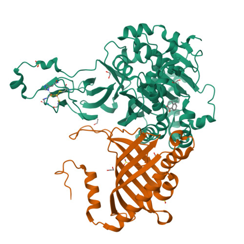
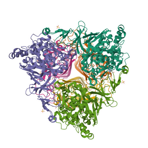
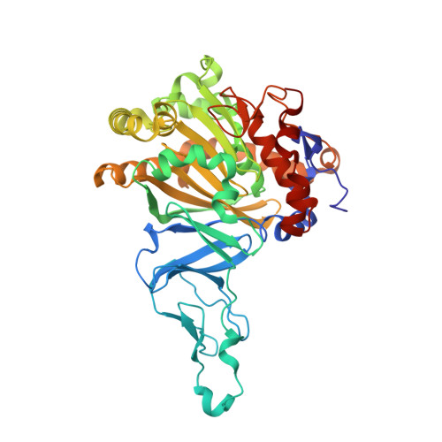
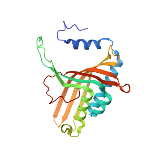
Crystal Structure of Naphthalene Dioxygenase: Side-on Binding of Dioxygen to Iron

Karlsson, A.Parales, J.V.Parales, R.E.Gibson, D.T.Eklund, H.Ramaswamy, S.

(2003) Science 299: 1039

  • DOI: https://doi.org/10.1126/science.1078020
  • Primary Citation of Related Structures:  
    1O7G, 1O7H, 1O7M, 1O7N, 1O7P, 1O7W

  • PubMed Abstract: 

    Binding of oxygen to iron is exploited in several biological and chemical processes. Although computational and spectroscopic results have suggested side-on binding, only end-on binding of oxygen to iron has been observed in crystal structures. We have determined structures of naphthalene dioxygenase that show a molecular oxygen species bound to the mononuclear iron in a side-on fashion. In a complex with substrate and dioxygen, the dioxygen molecule is lined up for an attack on the double bond of the aromatic substrate. The structures reported here provide the basis for a reaction mechanism and for the high stereospecificity of the reaction catalyzed by naphthalene dioxygenase.


  • Organizational Affiliation

    Department of Molecular Biology, Swedish University of Agricultural Sciences, Box 590, Biomedical Center, 75124 Uppsala, Sweden.


Macromolecules
Find similar proteins by:  (by identity cutoff)  |  3D Structure
Entity ID: 1
MoleculeChains Sequence LengthOrganismDetailsImage
NAPHTHALENE 1,2-DIOXYGENASE ALPHA SUBUNIT449Pseudomonas putidaMutation(s): 0 
EC: 1.14.12.12
UniProt
Find proteins for P0A110 (Pseudomonas putida)
Explore P0A110 
Go to UniProtKB:  P0A110
Entity Groups  
Sequence Clusters30% Identity50% Identity70% Identity90% Identity95% Identity100% Identity
UniProt GroupP0A110
Sequence Annotations
Expand
  • Reference Sequence
Find similar proteins by:  (by identity cutoff)  |  3D Structure
Entity ID: 2
MoleculeChains Sequence LengthOrganismDetailsImage
NAPHTHALENE 1,2-DIOXYGENASE BETA SUBUNIT194Pseudomonas putidaMutation(s): 0 
EC: 1.14.12.12
UniProt
Find proteins for P0A112 (Pseudomonas putida)
Explore P0A112 
Go to UniProtKB:  P0A112
Entity Groups  
Sequence Clusters30% Identity50% Identity70% Identity90% Identity95% Identity100% Identity
UniProt GroupP0A112
Sequence Annotations
Expand
  • Reference Sequence
Small Molecules
Ligands 5 Unique
IDChains Name / Formula / InChI Key2D Diagram3D Interactions
FES
Query on FES

Download Ideal Coordinates CCD File 
G [auth A]FE2/S2 (INORGANIC) CLUSTER
Fe2 S2
NIXDOXVAJZFRNF-UHFFFAOYSA-N
NDH
Query on NDH

Download Ideal Coordinates CCD File 
E [auth A](1R, 2S)-CIS 1,2 DIHYDROXY-1,2-DIHYDRONAPHTHALENE
C10 H10 O2
QPUHWUSUBHNZCG-VHSXEESVSA-N
SO4
Query on SO4

Download Ideal Coordinates CCD File 
F [auth A],
M [auth B],
N [auth B]
SULFATE ION
O4 S
QAOWNCQODCNURD-UHFFFAOYSA-L
EDO
Query on EDO

Download Ideal Coordinates CCD File 
C [auth A]
D [auth A]
I [auth B]
J [auth B]
K [auth B]
1,2-ETHANEDIOL
C2 H6 O2
LYCAIKOWRPUZTN-UHFFFAOYSA-N
FE
Query on FE

Download Ideal Coordinates CCD File 
H [auth A]FE (III) ION
Fe
VTLYFUHAOXGGBS-UHFFFAOYSA-N
Experimental Data & Validation

Experimental Data

  • Method: X-RAY DIFFRACTION
  • Resolution: 1.95 Å
  • R-Value Free:  0.219 (Depositor), 0.210 (DCC) 
  • R-Value Work:  0.184 (Depositor), 0.190 (DCC) 
Space Group: H 3 2
Unit Cell:
Length ( Å )Angle ( ˚ )
a = 139.673α = 90
b = 139.673β = 90
c = 208.439γ = 120
Software Package:
Software NamePurpose
REFMACrefinement
DENZOdata reduction
SCALEPACKdata scaling

Structure Validation

View Full Validation Report


Ligand Structure Quality Assessment 

Created with Raphaël 2.3.0Worse 01 BetterLigand structure goodness of fit to experimental dataBest fitted NDHClick on this verticalbar to view details

Entry History 

Deposition Data

Revision History  (Full details and data files)

  • Version 1.0: 2003-02-20
    Type: Initial release
  • Version 1.1: 2011-05-08
    Changes: Version format compliance
  • Version 1.2: 2011-07-13
    Changes: Version format compliance
  • Version 1.3: 2023-12-13
    Changes: Data collection, Database references, Derived calculations, Other, Refinement description