1YJD | pdb_00001yjd

Crystal structure of human CD28 in complex with the Fab fragment of a mitogenic antibody (5.11A1)


Experimental Data Snapshot

  • Method: X-RAY DIFFRACTION
  • Resolution: 2.70 Å
  • R-Value Free: 
    0.282 (Depositor), 0.285 (DCC) 
  • R-Value Work: 
    0.239 (Depositor), 0.242 (DCC) 
  • R-Value Observed: 
    0.248 (Depositor) 

wwPDB Validation   3D Report Full Report


Ligand Structure Quality Assessment 

Created with Raphaël 2.3.0Worse 01 BetterLigand structure goodness of fit to experimental dataBest fitted NAGClick on this verticalbar to view details

This is version 1.4 of the entry. See complete history


Literature

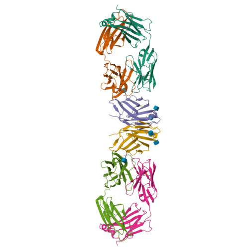
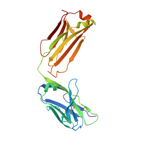
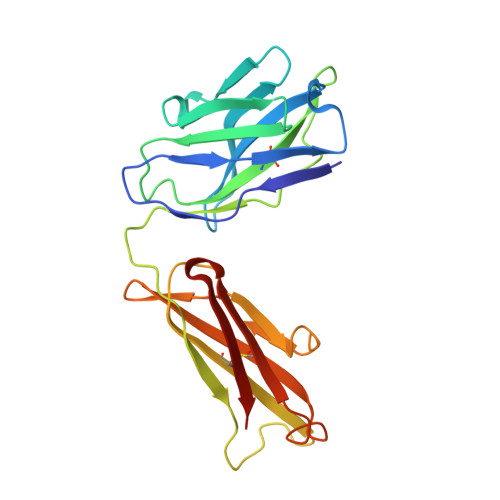
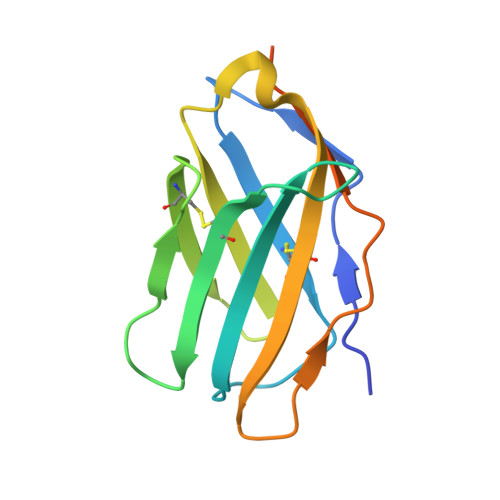
Crystal structure of a soluble CD28-Fab complex

Evans, E.J.Esnouf, R.M.Manso-Sancho, R.Gilbert, R.J.C.James, J.R.Yu, C.Fennelly, J.A.Vowles, C.Hanke, T.Walse, B.Hunig, T.Sorensen, P.Stuart, D.I.Davis, S.J.

(2005) Nat Immunol 6: 271-279

  • DOI: https://doi.org/10.1038/ni1170
  • Primary Citation of Related Structures:  
    1YJD

  • PubMed Abstract: 

    Naive T cell activation requires signaling by the T cell receptor and by nonclonotypic cell surface receptors. The most important costimulatory protein is the monovalent homodimer CD28, which interacts with CD80 and CD86 expressed on antigen-presenting cells. Here we present the crystal structure of a soluble form of CD28 in complex with the Fab fragment of a mitogenic antibody. Structural comparisons redefine the evolutionary relationships of CD28-related proteins, antigen receptors and adhesion molecules and account for the distinct ligand-binding and stoichiometric properties of CD28 and the related, inhibitory homodimer CTLA-4. Cryo-electron microscopy-based comparisons of complexes of CD28 with mitogenic and nonmitogenic antibodies place new constraints on models of antibody-induced receptor triggering. This work completes the initial structural characterization of the CD28-CTLA-4-CD80-CD86 signaling system.


  • Organizational Affiliation

    Nuffield Department of Clinical Medicine, The University of Oxford, John Radcliffe Hospital, Headington, Oxford, OX3 9DU, UK.


Macromolecules
Find similar proteins by:  (by identity cutoff)  |  3D Structure
Entity ID: 1
MoleculeChains Sequence LengthOrganismDetailsImage
Fab fragment of 5.11A1 antibody light chainA [auth L]212Mus musculusMutation(s): 0 
Entity Groups  
Sequence Clusters30% Identity50% Identity70% Identity90% Identity95% Identity100% Identity
Sequence Annotations
Expand
  • Reference Sequence
Find similar proteins by:  (by identity cutoff)  |  3D Structure
Entity ID: 2
MoleculeChains Sequence LengthOrganismDetailsImage
Fab fragment of 5.11A1 antibody heavy chainB [auth H]222Mus musculusMutation(s): 0 
Entity Groups  
Sequence Clusters30% Identity50% Identity70% Identity90% Identity95% Identity100% Identity
Sequence Annotations
Expand
  • Reference Sequence
Find similar proteins by:  (by identity cutoff)  |  3D Structure
Entity ID: 3
MoleculeChains Sequence LengthOrganismDetailsImage
T-cell-specific surface glycoprotein CD28140Homo sapiensMutation(s): 0 
Gene Names: CD28
UniProt & NIH Common Fund Data Resources
Find proteins for P10747 (Homo sapiens)
Explore P10747 
Go to UniProtKB:  P10747
PHAROS:  P10747
GTEx:  ENSG00000178562 
Entity Groups  
Sequence Clusters30% Identity50% Identity70% Identity90% Identity95% Identity100% Identity
UniProt GroupP10747
Glycosylation
Glycosylation Sites: 3Go to GlyGen: P10747-1
Sequence Annotations
Expand
  • Reference Sequence
Experimental Data & Validation

Experimental Data

  • Method: X-RAY DIFFRACTION
  • Resolution: 2.70 Å
  • R-Value Free:  0.282 (Depositor), 0.285 (DCC) 
  • R-Value Work:  0.239 (Depositor), 0.242 (DCC) 
  • R-Value Observed: 0.248 (Depositor) 
Space Group: C 1 2 1
Unit Cell:
Length ( Å )Angle ( ˚ )
a = 191.219α = 90
b = 47.417β = 94.45
c = 71.842γ = 90
Software Package:
Software NamePurpose
CNSrefinement
PDB_EXTRACTdata extraction
DENZOdata reduction
SCALEPACKdata scaling
X-PLORphasing

Structure Validation

View Full Validation Report


Ligand Structure Quality Assessment 

Created with Raphaël 2.3.0Worse 01 BetterLigand structure goodness of fit to experimental dataBest fitted NAGClick on this verticalbar to view details

Entry History 

Deposition Data

Revision History  (Full details and data files)

  • Version 1.0: 2005-02-15
    Type: Initial release
  • Version 1.1: 2008-04-30
    Changes: Version format compliance
  • Version 1.2: 2011-07-13
    Changes: Non-polymer description, Version format compliance
  • Version 1.3: 2020-07-29
    Type: Remediation
    Reason: Carbohydrate remediation
    Changes: Data collection, Database references, Derived calculations, Structure summary
  • Version 1.4: 2024-11-13
    Changes: Data collection, Database references, Structure summary