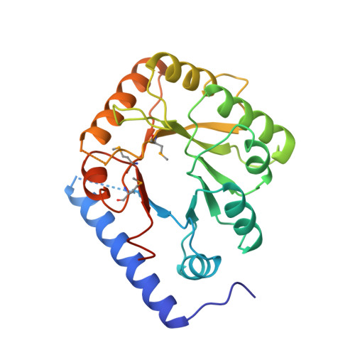
1CT5

CRYSTAL STRUCTURE OF YEAST HYPOTHETICAL PROTEIN YBL036C-SELENOMET CRYSTAL


Experimental Data Snapshot

  • Method: X-RAY DIFFRACTION
  • Resolution: 2.00 Å
  • R-Value Free: 0.245 
  • R-Value Work: 0.196 

wwPDB Validation   3D Report Full Report


Ligand Structure Quality Assessment 


This is version 1.3 of the entry. See complete history


Literature

Structure of a yeast hypothetical protein selected by a structural genomics approach.

Eswaramoorthy, S.Gerchman, S.Graziano, V.Kycia, H.Studier, F.W.Swaminathan, S.

(2003) Acta Crystallogr D Biol Crystallogr 59: 127-135

  • DOI: https://doi.org/10.1107/s0907444902018012
  • Primary Citation of Related Structures:  
    1B54, 1CT5

  • PubMed Abstract: 

    Yeast hypothetical protein YBL036C (SWISS-PROT P38197), initially thought to be a member of an 11-protein family, was selected for crystal structure determination since no structural or functional information was available. The structure has been determined independently by MIR and MAD methods to 2.0 A resolution. The MAD structure was determined largely through automated model building. The protein folds as a TIM barrel beginning with a long N-terminal helix, in contrast to the classic triose phosphate isomerase (TIM) structure, which begins with a beta-strand. A cofactor, pyridoxal 5'-phosphate, is covalently bound near the C-terminal end of the barrel, the usual active site in TIM-barrel folds. A single-domain monomeric molecule, this yeast protein resembles the N-terminal domain of alanine racemase or ornithine decarboxylase, both of which are two-domain dimeric proteins. The yeast protein has been shown to have amino-acid racemase activity. Although selected as a member of a protein family having no obvious relationship to proteins of known structure, the protein fold turned out to be a well known and widely distributed fold. This points to the need for a more comprehensive base of structural information and better structure-modeling tools before the goal of structure prediction from amino-acid sequences can be realised. In this case, similarity to a known structure allowed inferences to be made about the structure and function of a widely distributed protein family.


  • Organizational Affiliation

    Biology Department, Brookhaven National Laboratory, Upton, NY 11973, USA.


Macromolecules
Find similar proteins by:  (by identity cutoff)  |  3D Structure
Entity ID: 1
MoleculeChains Sequence LengthOrganismDetailsImage
PROTEIN (YEAST HYPOTHETICAL PROTEIN, SELENOMET)256Saccharomyces cerevisiaeMutation(s): 0 
UniProt
Find proteins for P38197 (Saccharomyces cerevisiae (strain ATCC 204508 / S288c))
Explore P38197 
Go to UniProtKB:  P38197
Entity Groups  
Sequence Clusters30% Identity50% Identity70% Identity90% Identity95% Identity100% Identity
UniProt GroupP38197
Sequence Annotations
Expand
  • Reference Sequence
Small Molecules
Ligands 1 Unique
IDChains Name / Formula / InChI Key2D Diagram3D Interactions
PLP
Query on PLP

Download Ideal Coordinates CCD File 
B [auth A]PYRIDOXAL-5'-PHOSPHATE
C8 H10 N O6 P
NGVDGCNFYWLIFO-UHFFFAOYSA-N
Modified Residues  1 Unique
IDChains TypeFormula2D DiagramParent
MSE
Query on MSE
A
L-PEPTIDE LINKINGC5 H11 N O2 SeMET
Experimental Data & Validation

Experimental Data

  • Method: X-RAY DIFFRACTION
  • Resolution: 2.00 Å
  • R-Value Free: 0.245 
  • R-Value Work: 0.196 
  • Space Group: P 21 21 21
Unit Cell:
Length ( Å )Angle ( ˚ )
a = 49.052α = 90
b = 65.823β = 90
c = 93.052γ = 90
Software Package:
Software NamePurpose
DENZOdata reduction
SCALEPACKdata scaling
PHASESphasing
SHARPphasing
REFMACrefinement

Structure Validation

View Full Validation Report


Ligand Structure Quality Assessment 


Entry History 

Deposition Data

Revision History  (Full details and data files)

  • Version 1.0: 1999-09-02
    Type: Initial release
  • Version 1.1: 2007-10-16
    Changes: Version format compliance
  • Version 1.2: 2011-07-13
    Changes: Non-polymer description, Version format compliance
  • Version 1.3: 2021-02-03
    Changes: Derived calculations, Structure summary