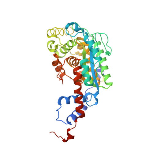
Structural investigation of the biosynthesis of alternative lower ligands for cobamides by nicotinate mononucleotide: 5,6-dimethylbenzimidazole phosphoribosyltransferase from Salmonella enterica.
Cheong, C.G., Escalante-Semerena, J.C., Rayment, I.(2001) J Biol Chem 276: 37612-37620
- PubMed: 11441022
- DOI: https://doi.org/10.1074/jbc.M105390200
- Primary Citation of Related Structures:
1JH8, 1JHA, 1JHM, 1JHO, 1JHP, 1JHQ, 1JHR, 1JHU, 1JHV, 1JHX, 1JHY - PubMed Abstract:
Nicotinate mononucleotide (NaMN):5,6-dimethylbenzimidazole phosphoribosyltransferase (CobT) from Salmonella enterica plays a central role in the synthesis of alpha-ribazole, a key component of the lower ligand of cobalamin. Surprisingly, CobT can phosphoribosylate a wide range of aromatic substrates, giving rise to a wide variety of lower ligands in cobamides. To understand the molecular basis for this lack of substrate specificity, the x-ray structures of CobT complexed with adenine, 5-methylbenzimidazole, 5-methoxybenzimidazole, p-cresol, and phenol were determined. Furthermore, adenine, 5-methylbenzimidazole, 5-methoxybenzimidazole, and 2-hydroxypurine were observed to react with NaMN within the crystal lattice and undergo the phosphoribosyl transfer reaction to form product. Significantly, the stereochemistries of all products are identical to those found in vivo. Interestingly, p-cresol and phenol, which are the lower ligand in Sporomusa ovata, bound to CobT but did not react with NaMN. This study provides a structural explanation for how CobT can phosphoribosylate most of the commonly observed lower ligands found in cobamides with the exception of the phenolic lower ligands observed in S. ovata. This is accomplished with minor conformational changes in the side chains that constitute the 5,6-dimethylbenzimidazole binding site. These investigations are consistent with the implication that the nature of the lower ligand is controlled by metabolic factors rather by the specificity of the phosphoribosyltransferase.
Organizational Affiliation:
Department of Biochemistry, University of Wisconsin, Madison, Wisconsin 53706, USA.


