1KJQ

Crystal structure of glycinamide ribonucleotide transformylase in complex with Mg-ADP


Experimental Data Snapshot

  • Method: X-RAY DIFFRACTION
  • Resolution: 1.05 Å
  • R-Value Free: 
    0.214 (Depositor) 
  • R-Value Work: 
    0.188 (Depositor), 0.190 (DCC) 
  • R-Value Observed: 
    0.190 (Depositor) 

Starting Model: experimental
View more details

wwPDB Validation   3D Report Full Report


Ligand Structure Quality Assessment 

Created with Raphaël 2.3.0Worse 01 BetterLigand structure goodness of fit to experimental dataBest fitted ADPClick on this verticalbar to view detailsBest fitted MPOClick on this verticalbar to view details

This is version 1.3 of the entry. See complete history


Literature

PurT-encoded glycinamide ribonucleotide transformylase. Accommodation of adenosine nucleotide analogs within the active site.

Thoden, J.B.Firestine, S.M.Benkovic, S.J.Holden, H.M.

(2002) J Biol Chem 277: 23898-23908

  • DOI: https://doi.org/10.1074/jbc.M202251200
  • Primary Citation of Related Structures:  
    1KJ8, 1KJ9, 1KJI, 1KJJ, 1KJQ

  • PubMed Abstract: 

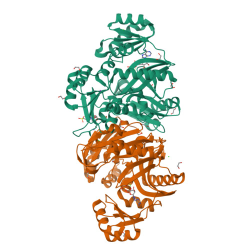
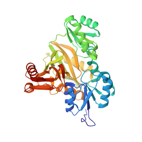
    PurT-encoded glycinamide ribonucleotide transformylase, or PurT transformylase, functions in purine biosynthesis by catalyzing the formylation of glycinamide ribonucleotide through a catalytic mechanism requiring Mg(2+)ATP and formate. From previous x-ray diffraction analyses, it has been demonstrated that PurT transformylase from Escherichia coli belongs to the ATP-grasp superfamily of enzymes, which are characterized by three structural motifs referred to as the A-, B-, and C-domains. In all of the ATP-grasp enzymes studied to date, the adenosine nucleotide ligands are invariably wedged between the B- and C-domains, and in some cases, such as biotin carboxylase and carbamoyl phosphate synthetase, the B-domains move significantly upon nucleotide binding. Here we present a systematic and high-resolution structural investigation of PurT transformylase complexed with various adenosine nucleotides or nucleotide analogs including Mg(2+)ATP, Mg(2+)-5'-adenylylimidodiphosphate, Mg(2+)-beta,gamma-methyleneadenosine 5'-triphosphate, Mg(2+)ATPgammaS, or Mg(2+)ADP. Taken together, these studies indicate that the conformation of the so-called "T-loop," delineated by Lys-155 to Gln-165, is highly sensitive to the chemical identity of the nucleotide situated in the binding pocket. This sensitivity to nucleotide identity is in sharp contrast to that observed for the "P-loop"-containing enzymes, in which the conformation of the binding motif is virtually unchanged in the presence or absence of nucleotides.


  • Organizational Affiliation

    Department of Biochemistry, University of Wisconsin, Madison, Wisconsin 53706, USA.


Macromolecules
Find similar proteins by:  (by identity cutoff)  |  3D Structure
Entity ID: 1
MoleculeChains Sequence LengthOrganismDetailsImage
phosphoribosylglycinamide formyltransferase 2
A, B
391Escherichia coliMutation(s): 0 
Gene Names: PURT
EC: 2.1.2 (PDB Primary Data), 6.3.1.21 (UniProt)
UniProt
Find proteins for P33221 (Escherichia coli (strain K12))
Explore P33221 
Go to UniProtKB:  P33221
Entity Groups  
Sequence Clusters30% Identity50% Identity70% Identity90% Identity95% Identity100% Identity
UniProt GroupP33221
Sequence Annotations
Expand
  • Reference Sequence
Small Molecules
Ligands 6 Unique
IDChains Name / Formula / InChI Key2D Diagram3D Interactions
ADP
Query on ADP

Download Ideal Coordinates CCD File 
G [auth A],
W [auth B]
ADENOSINE-5'-DIPHOSPHATE
C10 H15 N5 O10 P2
XTWYTFMLZFPYCI-KQYNXXCUSA-N
MPO
Query on MPO

Download Ideal Coordinates CCD File 
H [auth A]3[N-MORPHOLINO]PROPANE SULFONIC ACID
C7 H15 N O4 S
DVLFYONBTKHTER-UHFFFAOYSA-N
EDO
Query on EDO

Download Ideal Coordinates CCD File 
AA [auth B]
BA [auth B]
I [auth A]
J [auth A]
K [auth A]
1,2-ETHANEDIOL
C2 H6 O2
LYCAIKOWRPUZTN-UHFFFAOYSA-N
CL
Query on CL

Download Ideal Coordinates CCD File 
E [auth A],
F [auth A],
U [auth B],
V [auth B]
CHLORIDE ION
Cl
VEXZGXHMUGYJMC-UHFFFAOYSA-M
MG
Query on MG

Download Ideal Coordinates CCD File 
C [auth A],
S [auth B]
MAGNESIUM ION
Mg
JLVVSXFLKOJNIY-UHFFFAOYSA-N
NA
Query on NA

Download Ideal Coordinates CCD File 
D [auth A],
T [auth B]
SODIUM ION
Na
FKNQFGJONOIPTF-UHFFFAOYSA-N
Experimental Data & Validation

Experimental Data

  • Method: X-RAY DIFFRACTION
  • Resolution: 1.05 Å
  • R-Value Free:  0.214 (Depositor) 
  • R-Value Work:  0.188 (Depositor), 0.190 (DCC) 
  • R-Value Observed: 0.190 (Depositor) 
Space Group: P 21 21 2
Unit Cell:
Length ( Å )Angle ( ˚ )
a = 62.35α = 90
b = 179.34β = 90
c = 75.62γ = 90
Software Package:
Software NamePurpose
TNTrefinement
d*TREKdata reduction
HKL-2000data scaling
TNTphasing

Structure Validation

View Full Validation Report


Ligand Structure Quality Assessment 

Created with Raphaël 2.3.0Worse 01 BetterLigand structure goodness of fit to experimental dataBest fitted ADPClick on this verticalbar to view detailsBest fitted MPOClick on this verticalbar to view details

Entry History 

Deposition Data

Revision History  (Full details and data files)

  • Version 1.0: 2002-06-28
    Type: Initial release
  • Version 1.1: 2008-04-27
    Changes: Version format compliance
  • Version 1.2: 2011-07-13
    Changes: Version format compliance
  • Version 1.3: 2023-08-16
    Changes: Data collection, Database references, Derived calculations, Refinement description