1MQ5

Crystal Structure of 3-chloro-N-[4-chloro-2-[[(4-chlorophenyl)amino]carbonyl]phenyl]-4-[(4-methyl-1-piperazinyl)methyl]-2-thiophenecarboxamide Complexed with Human Factor Xa


Experimental Data Snapshot

  • Method: X-RAY DIFFRACTION
  • Resolution: 2.10 Å
  • R-Value Free: 0.264 
  • R-Value Work: 0.190 
  • R-Value Observed: 0.193 

Starting Model: experimental
View more details

wwPDB Validation   3D Report Full Report


Ligand Structure Quality Assessment 


This is version 1.4 of the entry. See complete history


Literature

Crystal Structures of Two Potent Nonamidine Inhibitors Bound to Factor Xa

Adler, M.Kochanny, M.J.Bin, Y.Rumennik, G.Light, D.L.Biancalana, S.Whitlow, M.

(2002) Biochemistry 41: 15514-15523

  • DOI: https://doi.org/10.1021/bi0264061
  • Primary Citation of Related Structures:  
    1MQ5, 1MQ6

  • PubMed Abstract: 

    There has been intense interest in the development of factor Xa inhibitors for the treatment of thrombotic diseases. Our laboratory has developed a series of novel non-amidine inhibitors of factor Xa. This paper presents two crystal structures of compounds from this series bound to factor Xa. The first structure is derived from the complex formed between factor Xa and compound 1. Compound 1 was the first non-amidine factor Xa inhibitor from our lab that had measurable potency in an in vitro assay of anticoagulant activity. The second compound, 2, has a molar affinity for factor Xa (K(iapp)) of 7 pM and good bioavailability. The two inhibitors bind in an L-shaped conformation with a chloroaromatic ring buried deeply in the S1 pocket. The opposite end of these compounds contains a basic substituent that extends into the S4 binding site. A chlorinated phenyl ring bridges the substituents in the S1 and S4 pockets via amide linkers. The overall conformation is similar to the previously published structures for amidine-based inhibitors complexed with factor Xa. However, there are significant differences in the interactions between the inhibitor and the protein at the atomic level. Most notably, there is no group that forms a salt bridge with the carboxylic acid at the base of the S1 pocket (Asp189). Each inhibitor forms only one well-defined hydrogen bond to the protein. There are no direct charge-charge interactions. The results indicate that electrostatic interactions play a secondary role in the binding of these potent inhibitors.


  • Organizational Affiliation

    Berlex Biosciences, 2600 Hilltop Drive, P.O. Box 4099, Richmond, CA 94804-0099, USA.


Macromolecules
Find similar proteins by:  (by identity cutoff)  |  3D Structure
Entity ID: 1
MoleculeChains Sequence LengthOrganismDetailsImage
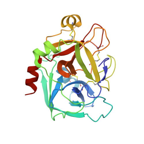
COAGULATION FACTOR X HEAVY CHAIN233Homo sapiensMutation(s): 0 
EC: 3.4.21.6
UniProt & NIH Common Fund Data Resources
Find proteins for P00742 (Homo sapiens)
Explore P00742 
Go to UniProtKB:  P00742
PHAROS:  P00742
GTEx:  ENSG00000126218 
Entity Groups  
Sequence Clusters30% Identity50% Identity70% Identity90% Identity95% Identity100% Identity
UniProt GroupP00742
Sequence Annotations
Expand
  • Reference Sequence
Find similar proteins by:  (by identity cutoff)  |  3D Structure
Entity ID: 2
MoleculeChains Sequence LengthOrganismDetailsImage
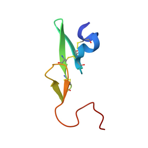
COAGULATION FACTOR X LIGHT CHAINB [auth L]51Homo sapiensMutation(s): 0 
EC: 3.4.21.6
UniProt & NIH Common Fund Data Resources
Find proteins for P00742 (Homo sapiens)
Explore P00742 
Go to UniProtKB:  P00742
PHAROS:  P00742
GTEx:  ENSG00000126218 
Entity Groups  
Sequence Clusters30% Identity50% Identity70% Identity90% Identity95% Identity100% Identity
UniProt GroupP00742
Sequence Annotations
Expand
  • Reference Sequence
Small Molecules
Binding Affinity Annotations 
IDSourceBinding Affinity
XLC BindingDB:  1MQ5 Ki: min: 0.76, max: 1 (nM) from 2 assay(s)
PDBBind:  1MQ5 Ki: 1 (nM) from 1 assay(s)
Experimental Data & Validation

Experimental Data

  • Method: X-RAY DIFFRACTION
  • Resolution: 2.10 Å
  • R-Value Free: 0.264 
  • R-Value Work: 0.190 
  • R-Value Observed: 0.193 
  • Space Group: P 21 21 21
Unit Cell:
Length ( Å )Angle ( ˚ )
a = 56.363α = 90
b = 72.018β = 90
c = 78.922γ = 90
Software Package:
Software NamePurpose
MAR345data collection
MOSFLMdata reduction
X-PLORmodel building
X-PLORrefinement
X-PLORphasing

Structure Validation

View Full Validation Report


Ligand Structure Quality Assessment 


Entry History 

Deposition Data

Revision History  (Full details and data files)

  • Version 1.0: 2003-01-28
    Type: Initial release
  • Version 1.1: 2008-04-28
    Changes: Version format compliance
  • Version 1.2: 2011-07-13
    Changes: Non-polymer description, Version format compliance
  • Version 1.3: 2017-10-11
    Changes: Advisory, Refinement description
  • Version 1.4: 2024-11-06
    Changes: Advisory, Data collection, Database references, Derived calculations, Refinement description, Structure summary