1MWR

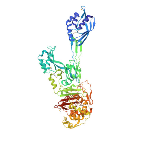
Structure of SeMet Penicillin binding protein 2a from methicillin resistant Staphylococcus aureus strain 27r (trigonal form) at 2.45 A resolution.


Experimental Data Snapshot

  • Method: X-RAY DIFFRACTION
  • Resolution: 2.45 Å
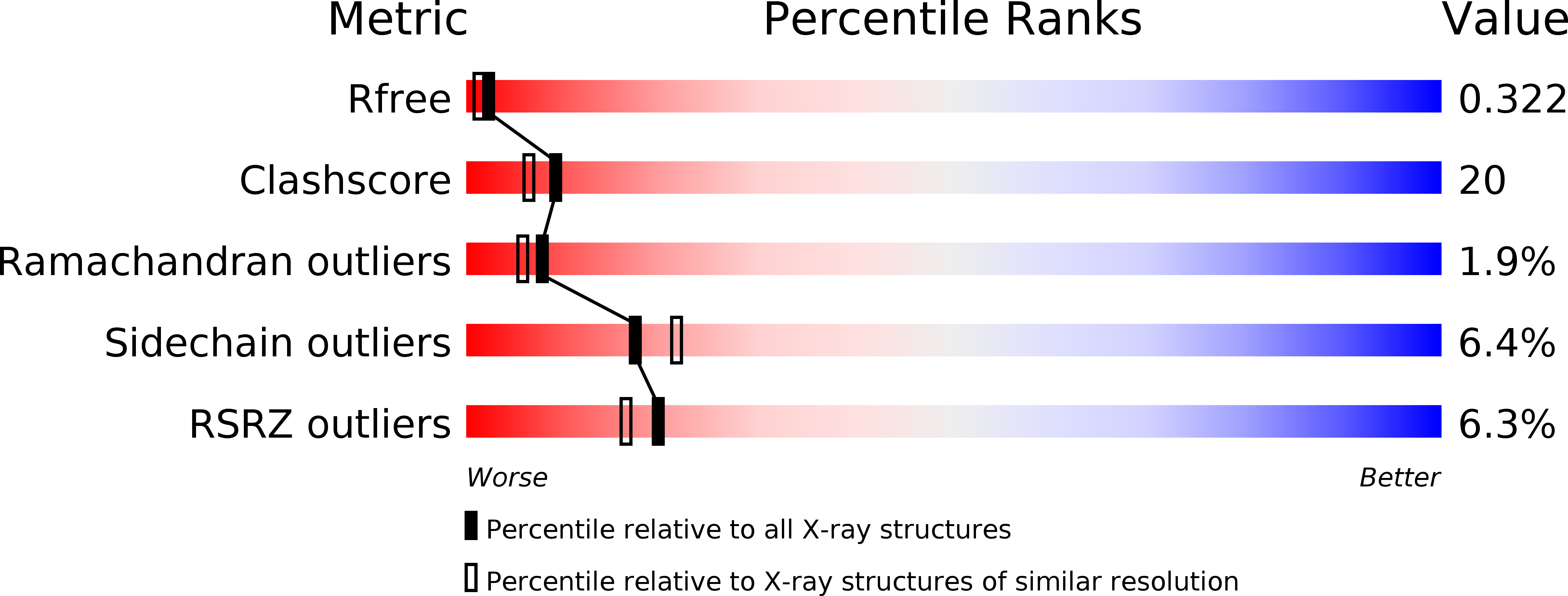
  • R-Value Free: 0.324 
  • R-Value Work: 0.270 
  • R-Value Observed: 0.270 

wwPDB Validation   3D Report Full Report


This is version 1.2 of the entry. See complete history


Literature

Structural basis for the beta lactam resistance of PBP2a from methicillin-resistant Staphylococcus aureus.

Lim, D.Strynadka, N.C.

(2002) Nat Struct Biol 9: 870-876

  • DOI: https://doi.org/10.1038/nsb858
  • Primary Citation of Related Structures:  
    1MWR, 1MWS, 1MWT, 1MWU, 1VQQ

  • PubMed Abstract: 

    The multiple antibiotic resistance of methicillin-resistant strains of Staphylococcus aureus (MRSA) has become a major clinical problem worldwide. The key determinant of the broad-spectrum beta-lactam resistance in MRSA strains is the penicillin-binding protein 2a (PBP2a). Because of its low affinity for beta-lactams, PBP2a provides transpeptidase activity to allow cell wall synthesis at beta-lactam concentrations that inhibit the beta-lactam-sensitive PBPs normally produced by S. aureus. The crystal structure of a soluble derivative of PBP2a has been determined to 1.8 A resolution and provides the highest resolution structure for a high molecular mass PBP. Additionally, structures of the acyl-PBP complexes of PBP2a with nitrocefin, penicillin G and methicillin allow, for the first time, a comparison of an apo and acylated resistant PBP. An analysis of the PBP2a active site in these forms reveals the structural basis of its resistance and identifies features in newly developed beta-lactams that are likely important for high affinity binding.


  • Organizational Affiliation

    2146 Health Sciences Mall, Department of Biochemistry and Molecular Biology, University of British Columbia, Vancouver, British Columbia V6T 1Z3, Canada.


Macromolecules
Find similar proteins by:  (by identity cutoff)  |  3D Structure
Entity ID: 1
MoleculeChains Sequence LengthOrganismDetailsImage
penicillin-binding protein 2a
A, B
646Staphylococcus aureusMutation(s): 0 
EC: 2.4.1.129
UniProt
Find proteins for Q93IC2 (Staphylococcus aureus)
Explore Q93IC2 
Go to UniProtKB:  Q93IC2
Entity Groups  
Sequence Clusters30% Identity50% Identity70% Identity90% Identity95% Identity100% Identity
UniProt GroupQ93IC2
Sequence Annotations
Expand
  • Reference Sequence
Small Molecules
Ligands 2 Unique
IDChains Name / Formula / InChI Key2D Diagram3D Interactions
CD
Query on CD

Download Ideal Coordinates CCD File 
C [auth A],
D [auth A],
G [auth B],
H [auth B]
CADMIUM ION
Cd
WLZRMCYVCSSEQC-UHFFFAOYSA-N
CL
Query on CL

Download Ideal Coordinates CCD File 
E [auth A],
F [auth A],
I [auth B],
J [auth B]
CHLORIDE ION
Cl
VEXZGXHMUGYJMC-UHFFFAOYSA-M
Modified Residues  1 Unique
IDChains TypeFormula2D DiagramParent
MSE
Query on MSE
A, B
L-PEPTIDE LINKINGC5 H11 N O2 SeMET
Experimental Data & Validation

Experimental Data

  • Method: X-RAY DIFFRACTION
  • Resolution: 2.45 Å
  • R-Value Free: 0.324 
  • R-Value Work: 0.270 
  • R-Value Observed: 0.270 
  • Space Group: P 31 2 1
Unit Cell:
Length ( Å )Angle ( ˚ )
a = 141.06α = 90
b = 141.06β = 90
c = 146.671γ = 120
Software Package:
Software NamePurpose
HKL-2000data collection
SCALEPACKdata scaling
SHARPphasing
CNSrefinement
HKL-2000data reduction

Structure Validation

View Full Validation Report


Entry History 

Deposition Data

Revision History  (Full details and data files)

  • Version 1.0: 2002-11-06
    Type: Initial release
  • Version 1.1: 2008-04-28
    Changes: Version format compliance
  • Version 1.2: 2011-07-13
    Changes: Version format compliance