1NF6

X-ray structure of the Desulfovibrio desulfuricans bacterioferritin: the diiron site in different catalytic states ("cycled" structure: reduced in solution and allowed to reoxidise before crystallisation)


Experimental Data Snapshot

  • Method: X-RAY DIFFRACTION
  • Resolution: 2.35 Å
  • R-Value Free: 
    0.267 (Depositor), 0.249 (DCC) 
  • R-Value Work: 
    0.197 (Depositor), 0.189 (DCC) 
  • R-Value Observed: 
    0.197 (Depositor) 

Starting Model: experimental
View more details

wwPDB Validation   3D Report Full Report


Ligand Structure Quality Assessment 

Created with Raphaël 2.3.0Worse 01 BetterLigand structure goodness of fit to experimental dataBest fitted FECClick on this verticalbar to view details

This is version 1.5 of the entry. See complete history


Literature

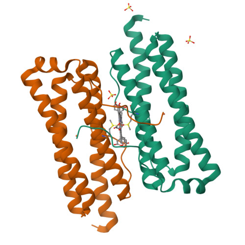
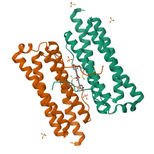
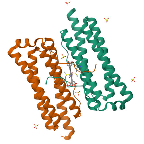
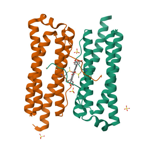
The nature of the di-iron site in the bacterioferritin from Desulfovibrio desulfuricans

Macedo, S.Romao, C.V.Mitchell, E.Matias, P.M.Liu, M.Y.Xavier, A.V.LeGall, J.Teixeira, M.Lindley, P.Carrondo, M.A.

(2003) Nat Struct Biol 10: 285-290

  • DOI: https://doi.org/10.1038/nsb909
  • Primary Citation of Related Structures:  
    1NF4, 1NF6, 1NFV

  • PubMed Abstract: 

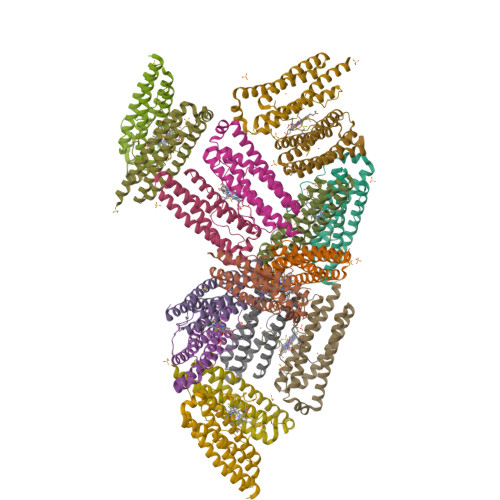
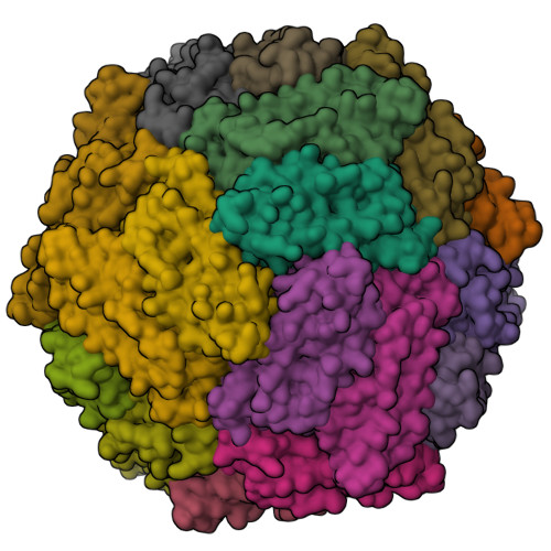
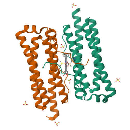
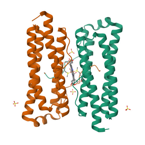
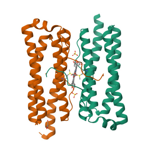
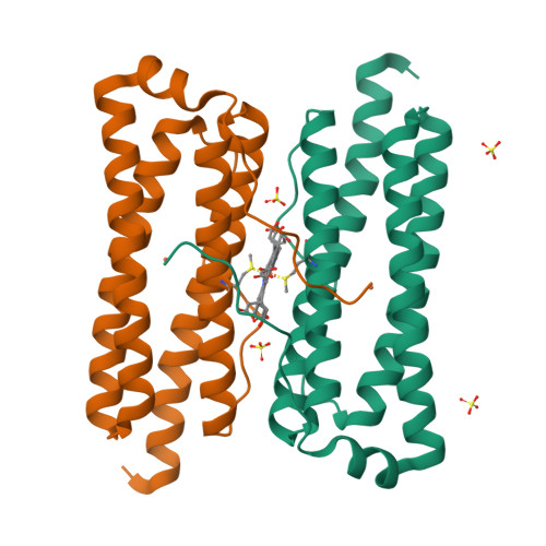
    The first crystal structure of a native di-iron center in an iron-storage protein (bacterio)ferritin is reported. The protein, isolated from the anaerobic bacterium Desulfovibrio desulfuricans, has the unique property of having Fe-coproporphyrin III as its heme cofactor. The three-dimensional structure of this bacterioferritin was determined in three distinct catalytic/redox states by X-ray crystallography (at 1.95, 2.05 and 2.35 A resolution), corresponding to different intermediates of the di-iron ferroxidase site. Conformational changes associated with these intermediates support the idea of a route for iron entry into the protein shell through a pore that passes through the di-iron center. Molecular surface and electrostatic potential calculations also suggest the presence of another ion channel, distant from the channels at the three- and four-fold axes proposed as points of entry for the iron atoms.


  • Organizational Affiliation

    Instituto de Tecnologia Química e Biológica, Universidade Nova de Lisboa, Av. República, EAN, Apartado 127, 2781-901 Oeiras, Portugal.


Macromolecules
Find similar proteins by:  (by identity cutoff)  |  3D Structure
Entity ID: 1
MoleculeChains Sequence LengthOrganismDetailsImage
bacterioferritin
A, B, C, D, E
179Desulfovibrio desulfuricansMutation(s): 0 
EC: 1.16.3.1
UniProt
Find proteins for Q93PP9 (Desulfovibrio desulfuricans (strain ATCC 27774 / DSM 6949 / MB))
Explore Q93PP9 
Go to UniProtKB:  Q93PP9
Entity Groups  
Sequence Clusters30% Identity50% Identity70% Identity90% Identity95% Identity100% Identity
UniProt GroupQ93PP9
Sequence Annotations
Expand
  • Reference Sequence
Small Molecules
Ligands 4 Unique
IDChains Name / Formula / InChI Key2D Diagram3D Interactions
FEC
Query on FEC

Download Ideal Coordinates CCD File 
AA [auth C]
EB [auth K]
JA [auth F]
NA [auth G]
QB [auth N]
1,3,5,8-TETRAMETHYL-PORPHINE-2,4,6,7-TETRAPROPIONIC ACID FERROUS COMPLEX
C36 H36 Fe N4 O8
FEDZMOFKVKOYTI-RGGAHWMASA-L
SO4
Query on SO4

Download Ideal Coordinates CCD File 
BB [auth K]
CA [auth D]
CB [auth K]
DA [auth E]
DB [auth K]
SULFATE ION
O4 S
QAOWNCQODCNURD-UHFFFAOYSA-L
GOL
Query on GOL

Download Ideal Coordinates CCD File 
WB [auth P]GLYCEROL
C3 H8 O3
PEDCQBHIVMGVHV-UHFFFAOYSA-N
FE
Query on FE

Download Ideal Coordinates CCD File 
AB [auth K]
BA [auth D]
EA [auth E]
FB [auth L]
GA [auth F]
FE (III) ION
Fe
VTLYFUHAOXGGBS-UHFFFAOYSA-N
Experimental Data & Validation

Experimental Data

  • Method: X-RAY DIFFRACTION
  • Resolution: 2.35 Å
  • R-Value Free:  0.267 (Depositor), 0.249 (DCC) 
  • R-Value Work:  0.197 (Depositor), 0.189 (DCC) 
  • R-Value Observed: 0.197 (Depositor) 
Space Group: P 21 3
Unit Cell:
Length ( Å )Angle ( ˚ )
a = 225.79α = 90
b = 225.79β = 90
c = 225.79γ = 90
Software Package:
Software NamePurpose
SHELXL-97refinement
SCALAdata scaling
CNSrefinement
DENZOdata reduction
CCP4data scaling
CNSphasing

Structure Validation

View Full Validation Report


Ligand Structure Quality Assessment 

Created with Raphaël 2.3.0Worse 01 BetterLigand structure goodness of fit to experimental dataBest fitted FECClick on this verticalbar to view details

Entry History 

Deposition Data

Revision History  (Full details and data files)

  • Version 1.0: 2003-04-01
    Type: Initial release
  • Version 1.1: 2008-04-29
    Changes: Version format compliance
  • Version 1.2: 2011-07-13
    Changes: Derived calculations, Version format compliance
  • Version 1.3: 2019-07-24
    Changes: Advisory, Data collection, Derived calculations, Refinement description
  • Version 1.4: 2024-04-03
    Changes: Advisory, Data collection, Database references, Derived calculations, Refinement description
  • Version 1.5: 2024-12-25
    Changes: Advisory, Derived calculations, Structure summary