1NHE

Crystal structure of Lactose synthase complex with UDP


Experimental Data Snapshot

  • Method: X-RAY DIFFRACTION
  • Resolution: 2.50 Å
  • R-Value Free: 0.255 
  • R-Value Work: 0.197 
  • R-Value Observed: 0.197 

wwPDB Validation   3D Report Full Report


Ligand Structure Quality Assessment 


This is version 1.3 of the entry. See complete history


Literature

Crystal structure of lactose synthase reveals a large conformational change in its catalytic component, the beta-1,4-galactosyltransferase

Ramakrishnan, B.Qasba, P.K.

(2001) J Mol Biol 310: 205-218

  • DOI: https://doi.org/10.1006/jmbi.2001.4757
  • Primary Citation of Related Structures:  
    1NF5, 1NHE, 1NKH, 1NQI

  • PubMed Abstract: 

    The lactose synthase (LS) enzyme is a 1:1 complex of a catalytic component, beta1,4-galactosyltransferse (beta4Gal-T1) and a regulatory component, alpha-lactalbumin (LA), a mammary gland-specific protein. LA promotes the binding of glucose (Glc) to beta4Gal-T1, thereby altering its sugar acceptor specificity from N-acetylglucosamine (GlcNAc) to glucose, which enables LS to synthesize lactose, the major carbohydrate component of milk. The crystal structures of LS bound with various substrates were solved at 2 A resolution. These structures reveal that upon substrate binding to beta4Gal-T1, a large conformational change occurs in the region comprising residues 345 to 365. This repositions His347 in such a way that it can participate in the coordination of a metal ion, and creates a sugar and LA-binding site. At the sugar-acceptor binding site, a hydrophobic N-acetyl group-binding pocket is found, formed by residues Arg359, Phe360 and Ile363. In the Glc-bound structure, this hydrophobic pocket is absent. For the binding of Glc to LS, a reorientation of the Arg359 side-chain occurs, which blocks the hydrophobic pocket and maximizes the interactions with the Glc molecule. Thus, the role of LA is to hold Glc by hydrogen bonding with the O-1 hydroxyl group in the acceptor-binding site on beta4Gal-T1, while the N-acetyl group-binding pocket in beta4Gal-T1 adjusts to maximize the interactions with the Glc molecule. This study provides details of a structural basis for the partially ordered kinetic mechanism proposed for lactose synthase.


  • Organizational Affiliation

    Structural Glycobiology Section, Intramural Research Support Program-SAIC, Laboratory of Experimental and Computational Biology, CCR, NCI, Frederick, MD 21702, USA.


Macromolecules
Find similar proteins by:  (by identity cutoff)  |  3D Structure
Entity ID: 1
MoleculeChains Sequence LengthOrganismDetailsImage
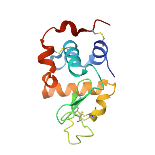
ALPHA-LACTALBUMIN
A, C
123Mus musculusMutation(s): 0 
UniProt
Find proteins for P29752 (Mus musculus)
Explore P29752 
Go to UniProtKB:  P29752
Entity Groups  
Sequence Clusters30% Identity50% Identity70% Identity90% Identity95% Identity100% Identity
UniProt GroupP29752
Sequence Annotations
Expand
  • Reference Sequence
Find similar proteins by:  (by identity cutoff)  |  3D Structure
Entity ID: 2
MoleculeChains Sequence LengthOrganismDetailsImage
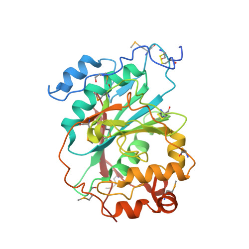
BETA-1,4-GALACTOSYLTRANSFERASE
B, D
286Bos taurusMutation(s): 0 
EC: 2.4.1.22 (PDB Primary Data), 2.4.1.90 (PDB Primary Data), 2.4.1.38 (PDB Primary Data), 2.4.1 (UniProt), 2.4.1.275 (UniProt)
UniProt
Find proteins for P08037 (Bos taurus)
Explore P08037 
Go to UniProtKB:  P08037
Entity Groups  
Sequence Clusters30% Identity50% Identity70% Identity90% Identity95% Identity100% Identity
UniProt GroupP08037
Sequence Annotations
Expand
  • Reference Sequence
Small Molecules
Modified Residues  1 Unique
IDChains TypeFormula2D DiagramParent
MSE
Query on MSE
B, D
L-PEPTIDE LINKINGC5 H11 N O2 SeMET
Experimental Data & Validation

Experimental Data

  • Method: X-RAY DIFFRACTION
  • Resolution: 2.50 Å
  • R-Value Free: 0.255 
  • R-Value Work: 0.197 
  • R-Value Observed: 0.197 
  • Space Group: P 1 21 1
Unit Cell:
Length ( Å )Angle ( ˚ )
a = 55.171α = 90
b = 99.013β = 103.92
c = 102.382γ = 90
Software Package:
Software NamePurpose
DENZOdata reduction
SCALEPACKdata scaling
SOLVEphasing
DMmodel building
CNSrefinement
DMphasing

Structure Validation

View Full Validation Report


Ligand Structure Quality Assessment 


Entry History 

Deposition Data

Revision History  (Full details and data files)

  • Version 1.0: 2003-01-07
    Type: Initial release
  • Version 1.1: 2008-04-29
    Changes: Version format compliance
  • Version 1.2: 2011-07-13
    Changes: Version format compliance
  • Version 1.3: 2024-10-30
    Changes: Data collection, Database references, Derived calculations, Structure summary