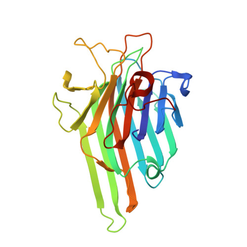
1NXD

Crystal structure of MnMn Concanavalin A


Experimental Data Snapshot

  • Method: X-RAY DIFFRACTION
  • Resolution: 1.90 Å
  • R-Value Free: 0.201 
  • R-Value Work: 0.184 

Starting Model: experimental
View more details

wwPDB Validation   3D Report Full Report


This is version 1.7 of the entry. See complete history


Literature

Structure of concanavalin A at pH 8: bound solvent and crystal contacts.

Lopez-Jaramillo, F.J.Gonzalez-Ramirez, L.A.Albert, A.Santoyo-Gonzalez, F.Vargas-Berenguel, A.Otalora, F.

(2004) Acta Crystallogr D Biol Crystallogr 60: 1048-1056

  • DOI: https://doi.org/10.1107/S0907444904007000
  • Primary Citation of Related Structures:  
    1NXD

  • PubMed Abstract: 

    Concanavalin A has been crystallized in the presence of the ligand (6-S-beta-D-galactopyranosyl-6-thio)-cyclomaltoheptaose. The crystals are isomorphous to those reported for ConA complexed with peptides at low resolution (3.00-2.75 angstroms). The structure was solved at 1.9 angstroms, with free R and R values of 0.201 and 0.184, respectively. As expected, no molecules of the ligand were bound to the protein. Soaking in the cryobuffer left its fingerprint as 25 molecules of glycerol in the bound solvent, most of them at specific positions. The fact that a glycerol molecule is located in the sugar-binding pocket of each of the four subunits in the asymmetric unit and another is located in two of the peptide-binding sites suggests a recognition phenomenon rather than a displacement of water molecules by glycerol. Crystal contact analysis shows that a relation exists between the residues that form hydrogen bonds to other asymmetric units and the space group: contact Asp58-Ser62 is a universal feature of ConA crystals, while Ser66-His121, Asn69-Asn118 and Tyr100-His205 contacts are general features of the C222(1) crystal form.


  • Organizational Affiliation

    Laboratorio de Estudios Cristalográficos, Instituto Andaluz de Ciencias de la Tierra, CSIC-UGRA, Facultad de Ciencias, Campus Fuentenueva, E-18002 Granada, Spain. javier@lec.ugr.es


Macromolecules
Find similar proteins by:  (by identity cutoff)  |  3D Structure
Entity ID: 1
MoleculeChains Sequence LengthOrganismDetailsImage
concanavalin AA [auth 1],
B [auth 2],
C [auth 3],
D [auth 4]
237Canavalia ensiformisMutation(s): 0 
UniProt
Find proteins for P02866 (Canavalia ensiformis)
Explore P02866 
Go to UniProtKB:  P02866
Entity Groups  
Sequence Clusters30% Identity50% Identity70% Identity90% Identity95% Identity100% Identity
UniProt GroupP02866
Sequence Annotations
Expand
  • Reference Sequence
Small Molecules
Ligands 4 Unique
IDChains Name / Formula / InChI Key2D Diagram3D Interactions
GOL
Query on GOL

Download Ideal Coordinates CCD File 
AA [auth 2]
AB [auth 4]
BA [auth 2]
BB [auth 4]
CA [auth 2]
AA [auth 2],
AB [auth 4],
BA [auth 2],
BB [auth 4],
CA [auth 2],
CB [auth 4],
DA [auth 2],
DB [auth 4],
EA [auth 2],
EB [auth 4],
FA [auth 2],
K [auth 1],
L [auth 1],
M [auth 1],
N [auth 1],
NA [auth 3],
O [auth 1],
OA [auth 3],
P [auth 1],
PA [auth 3],
Q [auth 1],
QA [auth 3],
R [auth 1],
RA [auth 3],
ZA [auth 4]
GLYCEROL
C3 H8 O3
PEDCQBHIVMGVHV-UHFFFAOYSA-N
MN
Query on MN

Download Ideal Coordinates CCD File 
H [auth 1]
I [auth 1]
KA [auth 3]
LA [auth 3]
WA [auth 4]
H [auth 1],
I [auth 1],
KA [auth 3],
LA [auth 3],
WA [auth 4],
X [auth 2],
XA [auth 4],
Y [auth 2]
MANGANESE (II) ION
Mn
WAEMQWOKJMHJLA-UHFFFAOYSA-N
AZI
Query on AZI

Download Ideal Coordinates CCD File 
E [auth 1]
F [auth 1]
G [auth 1]
GA [auth 3]
HA [auth 3]
E [auth 1],
F [auth 1],
G [auth 1],
GA [auth 3],
HA [auth 3],
IA [auth 3],
JA [auth 3],
S [auth 2],
SA [auth 4],
T [auth 2],
TA [auth 4],
U [auth 2],
UA [auth 4],
V [auth 2],
VA [auth 4],
W [auth 2]
AZIDE ION
N3
IVRMZWNICZWHMI-UHFFFAOYSA-N
NA
Query on NA

Download Ideal Coordinates CCD File 
J [auth 1],
MA [auth 3],
YA [auth 4],
Z [auth 2]
SODIUM ION
Na
FKNQFGJONOIPTF-UHFFFAOYSA-N
Experimental Data & Validation

Experimental Data

  • Method: X-RAY DIFFRACTION
  • Resolution: 1.90 Å
  • R-Value Free: 0.201 
  • R-Value Work: 0.184 
  • Space Group: C 2 2 21
Unit Cell:
Length ( Å )Angle ( ˚ )
a = 101.3α = 90
b = 118β = 90
c = 249.5γ = 90
Software Package:
Software NamePurpose
CNSrefinement
MAR345data collection
XDSdata scaling
AMoREphasing

Structure Validation

View Full Validation Report


Entry History 

Deposition Data

Revision History  (Full details and data files)

  • Version 1.0: 2004-03-30
    Type: Initial release
  • Version 1.1: 2008-04-29
    Changes: Version format compliance
  • Version 1.2: 2011-07-13
    Changes: Non-polymer description, Version format compliance
  • Version 1.3: 2011-11-16
    Changes: Atomic model
  • Version 1.4: 2017-07-05
    Changes: Source and taxonomy
  • Version 1.5: 2017-10-11
    Changes: Refinement description
  • Version 1.6: 2023-08-16
    Changes: Data collection, Database references, Derived calculations, Refinement description
  • Version 1.7: 2024-12-25
    Changes: Advisory, Derived calculations, Structure summary