Conformational Change in the NADP(H) Binding Domain of Transhydrogenase Defines Four States
Sundaresan, V., Yamaguchi, M., Chartron, J., Stout, C.D.(2003) Biochemistry 42: 12143-12153
- PubMed: 14567675
- DOI: https://doi.org/10.1021/bi035006q
- Primary Citation of Related Structures:
1PNO, 1PNQ - PubMed Abstract:
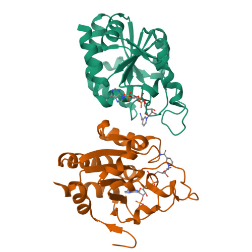
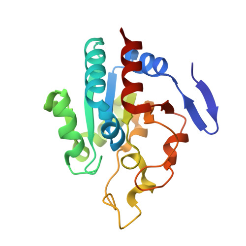
Proton-translocating transhydrogenase (TH) couples direct and stereospecific hydride transfer between NAD(H) and NADP(H), bound to soluble domains dI and dIII, respectively, to proton translocation across a membrane bound domain, dII. The reaction occurs with proton-gradient coupled conformational changes, which affect the energetics of substrate binding and interdomain interactions. The crystal structure of TH dIII from Rhodospirillum rubrum has been determined in the presence of NADPH (2.4 A) and NADP (2.1 A) (space group P6(1)22). Each structure has two molecules in the asymmetric unit, differing in the conformation of the NADP(H) binding loop D. In one molecule, loop D has an open conformation, with the B face of (dihydro)nicotinamide exposed to solvent. In the other molecule, loop D adopts a hitherto unobserved closed conformation, resulting in close interactions between NADP(H) and side chains of the highly conserved residues, betaSer405, betaPro406, and betaIle407. The conformational change shields the B face of (dihydro)nicotinamide from solvent, which would block hydride transfer in the intact enzyme. It also alters the environments of invariant residues betaHis346 and betaAsp393. However, there is little difference in either the open or the closed conformation upon change in oxidation state of nicotinamide, i.e., for NADP vs. NADPH. Consequently, the occurrence of two loop D conformations for both substrate oxidation states gives rise to four states: NADP-open, NADP-closed, NADPH-open, and NADPH-closed. Because these states are distinguished by protein conformation and by net charge they may be important in the proton translocating mechanism of intact TH.
Organizational Affiliation:
Department of Molecular Biology, 10550 North Torrey Pines Road, The Scripps Research Institute, La Jolla, California 92037, USA.


