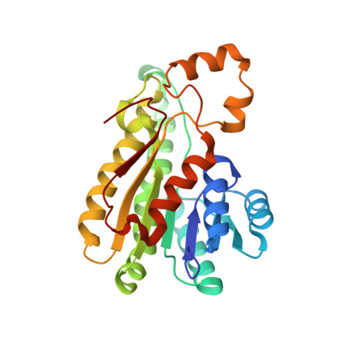
1Q7B

The structure of betaketoacyl-[ACP] reductase from E. coli in complex with NADP+


Experimental Data Snapshot

  • Method: X-RAY DIFFRACTION
  • Resolution: 2.05 Å
  • R-Value Free: 0.221 
  • R-Value Work: 0.200 
  • R-Value Observed: 0.201 

wwPDB Validation   3D Report Full Report


Ligand Structure Quality Assessment 


This is version 1.3 of the entry. See complete history


Literature

Cofactor-Induced Conformational Rearrangements Establish a Catalytically Competent Active Site and a Proton Relay Conduit in FabG

Price, A.C.Zhang, Y.-M.Rock, C.O.White, S.M.

(2004) Structure 12: 417-428

  • DOI: https://doi.org/10.1016/j.str.2004.02.008
  • Primary Citation of Related Structures:  
    1Q7B, 1Q7C

  • PubMed Abstract: 

    beta-Ketoacyl-acyl carrier protein reductase (FabG) is a key component in the type II fatty acid synthase system. The structures of Escherichia coli FabG and the FabG[Y151F] mutant in binary complexes with NADP(H) reveal that mechanistically important conformational changes accompany cofactor binding. The active site Ser-Tyr-Lys triad is repositioned into a catalytically competent constellation, and a hydrogen bonded network consisting of ribose hydroxyls, the Ser-Tyr-Lys triad, and four water molecules creates a proton wire to replenish the tyrosine proton donated during catalysis. Also, a disordered loop in FabG forms a substructure in the complex that shapes the entrance to the active site. A key observation is that the nicotinamide portion of the cofactor is disordered in the FabG[Y151F].NADP(H) complex, and Tyr151 appears to be necessary for high-affinity cofactor binding. Biochemical data confirm that FabG[Y151F] is defective in NADPH binding. Finally, structural changes consistent with the observed negative cooperativity of FabG are described.


  • Organizational Affiliation

    Department of Structural Biology, St Jude Children's Research Hospital, Memphis, TN 38105 USA.


Macromolecules
Find similar proteins by:  (by identity cutoff)  |  3D Structure
Entity ID: 1
MoleculeChains Sequence LengthOrganismDetailsImage
3-oxoacyl-[acyl-carrier protein] reductase
A, B, C, D
244Escherichia coliMutation(s): 0 
EC: 1.1.1.100
UniProt
Find proteins for P0AEK2 (Escherichia coli (strain K12))
Explore P0AEK2 
Go to UniProtKB:  P0AEK2
Entity Groups  
Sequence Clusters30% Identity50% Identity70% Identity90% Identity95% Identity100% Identity
UniProt GroupP0AEK2
Sequence Annotations
Expand
  • Reference Sequence
Small Molecules
Ligands 2 Unique
IDChains Name / Formula / InChI Key2D Diagram3D Interactions
NAP
Query on NAP

Download Ideal Coordinates CCD File 
H [auth A],
L [auth B],
N [auth C],
P [auth D]
NADP NICOTINAMIDE-ADENINE-DINUCLEOTIDE PHOSPHATE
C21 H28 N7 O17 P3
XJLXINKUBYWONI-NNYOXOHSSA-N
CA
Query on CA

Download Ideal Coordinates CCD File 
E [auth A]
F [auth A]
G [auth A]
I [auth B]
J [auth B]
E [auth A],
F [auth A],
G [auth A],
I [auth B],
J [auth B],
K [auth B],
M [auth C],
O [auth D]
CALCIUM ION
Ca
BHPQYMZQTOCNFJ-UHFFFAOYSA-N
Experimental Data & Validation

Experimental Data

  • Method: X-RAY DIFFRACTION
  • Resolution: 2.05 Å
  • R-Value Free: 0.221 
  • R-Value Work: 0.200 
  • R-Value Observed: 0.201 
  • Space Group: P 1 21 1
Unit Cell:
Length ( Å )Angle ( ˚ )
a = 72.748α = 90
b = 76.182β = 109.47
c = 99.733γ = 90
Software Package:
Software NamePurpose
DENZOdata reduction
SCALEPACKdata scaling
AMoREphasing
CNSrefinement

Structure Validation

View Full Validation Report


Ligand Structure Quality Assessment 


Entry History 

Deposition Data

Revision History  (Full details and data files)

  • Version 1.0: 2004-02-17
    Type: Initial release
  • Version 1.1: 2008-04-29
    Changes: Version format compliance
  • Version 1.2: 2011-07-13
    Changes: Version format compliance
  • Version 1.3: 2024-02-14
    Changes: Data collection, Database references, Derived calculations