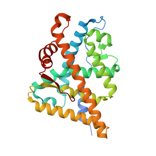
1T79

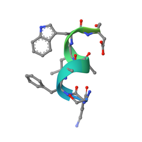
Crystal structure of the androgen receptor ligand binding domain in complex with a FxxLW motif


Experimental Data Snapshot

  • Method: X-RAY DIFFRACTION
  • Resolution: 1.80 Å
  • R-Value Free: 0.234 
  • R-Value Work: 0.200 

wwPDB Validation   3D Report Full Report


Ligand Structure Quality Assessment 


This is version 1.3 of the entry. See complete history


Literature

Recognition and accommodation at the androgen receptor coactivator binding interface.

Hur, E.Pfaff, S.J.Payne, E.S.Gron, H.Buehrer, B.M.Fletterick, R.J.

(2004) PLoS Biol 2: E274-E274

  • DOI: https://doi.org/10.1371/journal.pbio.0020274
  • Primary Citation of Related Structures:  
    1T73, 1T74, 1T76, 1T79, 1T7F, 1T7M, 1T7R, 1T7T

  • PubMed Abstract: 

    Prostate cancer is a leading killer of men in the industrialized world. Underlying this disease is the aberrant action of the androgen receptor (AR). AR is distinguished from other nuclear receptors in that after hormone binding, it preferentially responds to a specialized set of coactivators bearing aromatic-rich motifs, while responding poorly to coactivators bearing the leucine-rich "NR box" motifs favored by other nuclear receptors. Under normal conditions, interactions with these AR-specific coactivators through aromatic-rich motifs underlie targeted gene transcription. However, during prostate cancer, abnormal association with such coactivators, as well as with coactivators containing canonical leucine-rich motifs, promotes disease progression. To understand the paradox of this unusual selectivity, we have derived a complete set of peptide motifs that interact with AR using phage display. Binding affinities were measured for a selected set of these peptides and their interactions with AR determined by X-ray crystallography. Structures of AR in complex with FxxLF, LxxLL, FxxLW, WxxLF, WxxVW, FxxFF, and FxxYF motifs reveal a changing surface of the AR coactivator binding interface that permits accommodation of both AR-specific aromatic-rich motifs and canonical leucine-rich motifs. Induced fit provides perfect mating of the motifs representing the known family of AR coactivators and suggests a framework for the design of AR coactivator antagonists.


  • Organizational Affiliation

    Graduate Group in Biophysics, University of California, San Francisco, California, USA.


Macromolecules
Find similar proteins by:  (by identity cutoff)  |  3D Structure
Entity ID: 1
MoleculeChains Sequence LengthOrganismDetailsImage
Androgen receptor269Pan troglodytesMutation(s): 0 
Gene Names: ARNR3C4
UniProt
Find proteins for O97775 (Pan troglodytes)
Explore O97775 
Go to UniProtKB:  O97775
Entity Groups  
Sequence Clusters30% Identity50% Identity70% Identity90% Identity95% Identity100% Identity
UniProt GroupO97775
Sequence Annotations
Expand
  • Reference Sequence

Find similar proteins by:  Sequence   |   3D Structure  

Entity ID: 2
MoleculeChains Sequence LengthOrganismDetailsImage
FxxLW motif peptide20N/AMutation(s): 0 
Entity Groups  
Sequence Clusters30% Identity50% Identity70% Identity90% Identity95% Identity100% Identity
Sequence Annotations
Expand
  • Reference Sequence
Small Molecules
Ligands 1 Unique
IDChains Name / Formula / InChI Key2D Diagram3D Interactions
DHT
Query on DHT

Download Ideal Coordinates CCD File 
C [auth A]5-ALPHA-DIHYDROTESTOSTERONE
C19 H30 O2
NVKAWKQGWWIWPM-ABEVXSGRSA-N
Binding Affinity Annotations 
IDSourceBinding Affinity
DHT BindingDB:  1T79 Ki: min: 0.2, max: 10 (nM) from 12 assay(s)
Kd: 0.25 (nM) from 1 assay(s)
IC50: min: 0.05, max: 18.5 (nM) from 14 assay(s)
EC50: min: 0.05, max: 20 (nM) from 16 assay(s)
Experimental Data & Validation

Experimental Data

  • Method: X-RAY DIFFRACTION
  • Resolution: 1.80 Å
  • R-Value Free: 0.234 
  • R-Value Work: 0.200 
  • Space Group: P 21 21 21
Unit Cell:
Length ( Å )Angle ( ˚ )
a = 55.826α = 90
b = 66.128β = 90
c = 68.381γ = 90
Software Package:
Software NamePurpose
CNSrefinement
DENZOdata reduction
SCALEPACKdata scaling
CNSphasing

Structure Validation

View Full Validation Report


Ligand Structure Quality Assessment 


Entry History 

Deposition Data

Revision History  (Full details and data files)

  • Version 1.0: 2004-08-31
    Type: Initial release
  • Version 1.1: 2008-04-30
    Changes: Version format compliance
  • Version 1.2: 2011-07-13
    Changes: Version format compliance
  • Version 1.3: 2024-02-14
    Changes: Data collection, Database references, Derived calculations