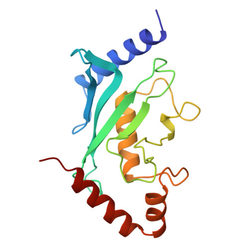
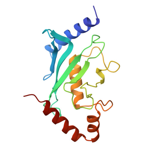
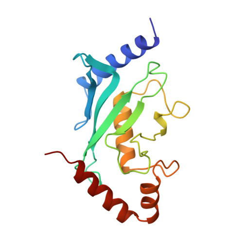
1U9A

HUMAN UBIQUITIN-CONJUGATING ENZYME UBC9


Experimental Data Snapshot

  • Method: X-RAY DIFFRACTION
  • Resolution: 2.00 Å
  • R-Value Free: 0.250 
  • R-Value Work: 0.160 

Starting Model: experimental
View more details

wwPDB Validation   3D Report Full Report


This is version 1.4 of the entry. See complete history


Literature

Crystal structure of murine/human Ubc9 provides insight into the variability of the ubiquitin-conjugating system.

Tong, H.Hateboer, G.Perrakis, A.Bernards, R.Sixma, T.K.

(1997) J Biol Chem 272: 21381-21387

  • DOI: https://doi.org/10.1074/jbc.272.34.21381
  • Primary Citation of Related Structures:  
    1U9A, 1U9B

  • PubMed Abstract: 

    Murine/human ubiquitin-conjugating enzyme Ubc9 is a functional homolog of Saccharomyces cerevisiae Ubc9 that is essential for the viability of yeast cells with a specific role in the G2-M transition of the cell cycle. The structure of recombinant mammalian Ubc9 has been determined from two crystal forms at 2.0 A resolution. Like Arabidopsis thaliana Ubc1 and S. cerevisiae Ubc4, murine/human Ubc9 was crystallized as a monomer, suggesting that previously reported hetero- and homo-interactions among Ubcs may be relatively weak or indirect. Compared with the known crystal structures of Ubc1 and Ubc4, which regulate different cellular processes, Ubc9 has a 5-residue insertion that forms a very exposed tight beta-hairpin and a 2-residue insertion that forms a bulge in a loop close to the active site. Mammalian Ubc9 also possesses a distinct electrostatic potential distribution that may provide possible clues to its remarkable ability to interact with other proteins. The 2-residue insertion and other sequence and structural heterogeneity observed at the catalytic site suggest that different Ubcs may utilize catalytic mechanisms of varying efficiency and substrate specificity.


  • Organizational Affiliation

    Netherlands Cancer Institute, Plesmanlaan 121, 1066 CX, Amsterdam, The Netherlands.


Macromolecules
Find similar proteins by:  (by identity cutoff)  |  3D Structure
Entity ID: 1
MoleculeChains Sequence LengthOrganismDetailsImage
UBIQUITIN-CONJUGATING ENZYME160Mus musculusMutation(s): 0 
EC: 6.3.2.19 (PDB Primary Data), 2.3.2 (UniProt)
UniProt & NIH Common Fund Data Resources
Find proteins for P63280 (Mus musculus)
Explore P63280 
Go to UniProtKB:  P63280
IMPC:  MGI:107365
Entity Groups  
Sequence Clusters30% Identity50% Identity70% Identity90% Identity95% Identity100% Identity
UniProt GroupP63280
Sequence Annotations
Expand
  • Reference Sequence
Experimental Data & Validation

Experimental Data

  • Method: X-RAY DIFFRACTION
  • Resolution: 2.00 Å
  • R-Value Free: 0.250 
  • R-Value Work: 0.160 
  • Space Group: P 1 21 1
Unit Cell:
Length ( Å )Angle ( ˚ )
a = 52.04α = 90
b = 35.18β = 111.2
c = 58.1γ = 90
Software Package:
Software NamePurpose
AMoREphasing
TNTrefinement
DENZOdata reduction
SCALEPACKdata scaling

Structure Validation

View Full Validation Report


Entry History 

Deposition Data

Revision History  (Full details and data files)

  • Version 1.0: 1997-05-15
    Type: Initial release
  • Version 1.1: 2008-03-24
    Changes: Version format compliance
  • Version 1.2: 2011-07-13
    Changes: Version format compliance
  • Version 1.3: 2023-08-09
    Changes: Data collection, Database references, Refinement description
  • Version 1.4: 2024-05-22
    Changes: Data collection