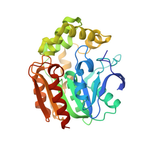
1UK8

Crystal structure of a meta-cleavage product hydrolase (CumD) complexed with n-valerate


Experimental Data Snapshot

  • Method: X-RAY DIFFRACTION
  • Resolution: 1.60 Å
  • R-Value Free: 0.190 
  • R-Value Work: 0.178 
  • R-Value Observed: 0.178 

Starting Model: experimental
View more details

wwPDB Validation   3D Report Full Report


This is version 1.4 of the entry. See complete history


Literature

A Series of Crystal Structures of a meta-Cleavage Product Hydrolase from Pseudomonas fluorescens IP01 (CumD) Complexed with Various Cleavage Products

Fushinobu, S.Jun, S.-Y.Hidaka, M.Nojiri, H.Yamane, H.Shoun, H.Omori, T.Wakagi, T.

(2005) Biosci Biotechnol Biochem 69: 491-498

  • DOI: https://doi.org/10.1271/bbb.69.491
  • Primary Citation of Related Structures:  
    1UK6, 1UK7, 1UK8, 1UK9, 1UKA, 1UKB

  • PubMed Abstract: 

    Meta-cleavage product hydrolase (MCP-hydrolase) is one of the key enzymes in the microbial degradation of aromatic compounds. MCP-hydrolase produces 2-hydroxypenta-2,4-dienoate and various organic acids, according to the C6 substituent of the substrate. Comprehensive analysis of the substrate specificity of the MCP-hydrolase from Pseudomonas fluorescens IP01 (CumD) was carried out by determining the kinetic parameters for nine substrates and crystal structures complexed with eight cleavage products. CumD preferred substrates with long non-branched C6 substituents, but did not effectively hydrolyze a substrate with a phenyl group. Superimposition of the complex structures indicated that benzoate was bound in a significantly different direction than other aliphatic cleavage products. The directions of the bound organic acids appeared to be related with the k(cat) values of the corresponding substrates. The Ile139 and Trp143 residues on helix alpha4 appeared to cause steric hindrance with the aromatic ring of the substrate, which hampers base-catalyzed attack by water.


  • Organizational Affiliation

    Department of Biotechnology, The University of Tokyo, Japan. asfushi@mail.ecc.u-tokyo.ac.jp


Macromolecules
Find similar proteins by:  (by identity cutoff)  |  3D Structure
Entity ID: 1
MoleculeChains Sequence LengthOrganismDetailsImage
2-hydroxy-6-oxo-7-methylocta-2,4-dienoate hydrolase282Pseudomonas fluorescensMutation(s): 1 
Gene Names: CUMD
EC: 3.7.1.9
UniProt
Find proteins for P96965 (Pseudomonas fluorescens)
Explore P96965 
Go to UniProtKB:  P96965
Entity Groups  
Sequence Clusters30% Identity50% Identity70% Identity90% Identity95% Identity100% Identity
UniProt GroupP96965
Sequence Annotations
Expand
  • Reference Sequence
Experimental Data & Validation

Experimental Data

  • Method: X-RAY DIFFRACTION
  • Resolution: 1.60 Å
  • R-Value Free: 0.190 
  • R-Value Work: 0.178 
  • R-Value Observed: 0.178 
  • Space Group: C 2 2 21
Unit Cell:
Length ( Å )Angle ( ˚ )
a = 77.101α = 90
b = 116.001β = 90
c = 78.73γ = 90
Software Package:
Software NamePurpose
CNSrefinement
MOSFLMdata reduction
CCP4data scaling
CNSphasing

Structure Validation

View Full Validation Report


Entry History 

Deposition Data

Revision History  (Full details and data files)

  • Version 1.0: 2004-09-14
    Type: Initial release
  • Version 1.1: 2008-04-27
    Changes: Version format compliance
  • Version 1.2: 2011-07-13
    Changes: Version format compliance
  • Version 1.3: 2021-11-10
    Changes: Database references, Derived calculations
  • Version 1.4: 2023-10-25
    Changes: Data collection, Refinement description