1Y9M | pdb_00001y9m

Crystal structure of exo-inulinase from Aspergillus awamori in spacegroup P212121


Experimental Data Snapshot

  • Method: X-RAY DIFFRACTION
  • Resolution: 1.89 Å
  • R-Value Free: 
    0.200 (Depositor), 0.199 (DCC) 
  • R-Value Work: 
    0.165 (Depositor), 0.165 (DCC) 
  • R-Value Observed: 
    0.167 (Depositor) 

wwPDB Validation   3D Report Full Report


Ligand Structure Quality Assessment 

Created with Raphaël 2.3.0Worse 01 BetterLigand structure goodness of fit to experimental dataBest fitted NAGClick on this verticalbar to view detailsBest fitted MANClick on this verticalbar to view details

This is version 2.1 of the entry. See complete history


Literature

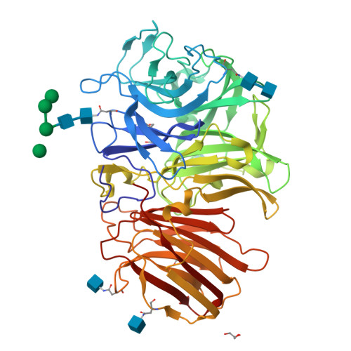
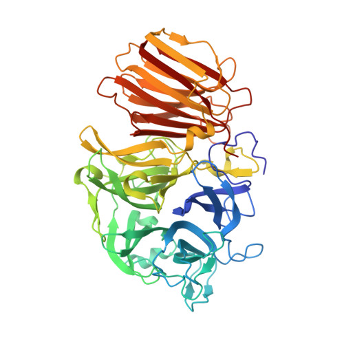
Crystal structure of exo-inulinase from Aspergillus awamori: the enzyme fold and structural determinants of substrate recognition

Nagem, R.A.P.Rojas, A.L.Golubev, A.M.Korneeva, O.S.Eneyskaya, E.V.Kulminskaya, A.A.Neustroev, K.N.Polikarpov, I.

(2004) J Mol Biology 344: 471-480

  • DOI: https://doi.org/10.1016/j.jmb.2004.09.024
  • Primary Citation of Related Structures:  
    1Y4W, 1Y9G, 1Y9M

  • PubMed Abstract: 

    Exo-inulinases hydrolyze terminal, non-reducing 2,1-linked and 2,6-linked beta-d-fructofuranose residues in inulin, levan and sucrose releasing beta-d-fructose. We present the X-ray structure at 1.55A resolution of exo-inulinase from Aspergillus awamori, a member of glycoside hydrolase family 32, solved by single isomorphous replacement with the anomalous scattering method using the heavy-atom sites derived from a quick cryo-soaking technique. The tertiary structure of this enzyme folds into two domains: the N-terminal catalytic domain of an unusual five-bladed beta-propeller fold and the C-terminal domain folded into a beta-sandwich-like structure. Its structural architecture is very similar to that of another member of glycoside hydrolase family 32, invertase (beta-fructosidase) from Thermotoga maritima, determined recently by X-ray crystallography The exo-inulinase is a glycoprotein containing five N-linked oligosaccharides. Two crystal forms obtained under similar crystallization conditions differ by the degree of protein glycosylation. The X-ray structure of the enzyme:fructose complex, at a resolution of 1.87A, reveals two catalytically important residues: Asp41 and Glu241, a nucleophile and a catalytic acid/base, respectively. The distance between the side-chains of these residues is consistent with a double displacement mechanism of reaction. Asp189, which is part of the Arg-Asp-Pro motif, provides hydrogen bonds important for substrate recognition.


  • Organizational Affiliation

    Instituto de Física de São Carlos, Universidade de São Paulo, Av. Trabalhador São-carlense 400, CEP 13560-970, São Carlos, SP, Brazil.


Macromolecules
Find similar proteins by:  (by identity cutoff)  |  3D Structure
Entity ID: 1
MoleculeChains Sequence LengthOrganismDetailsImage
exo-inulinase518Aspergillus awamoriMutation(s): 0 
EC: 3.2.1.80
UniProt
Find proteins for Q96TU3 (Aspergillus awamori)
Explore Q96TU3 
Go to UniProtKB:  Q96TU3
Entity Groups  
Sequence Clusters30% Identity50% Identity70% Identity90% Identity95% Identity100% Identity
UniProt GroupQ96TU3
Glycosylation
Glycosylation Sites: 5
Sequence Annotations
Expand
  • Reference Sequence
Oligosaccharides

Help

Entity ID: 2
MoleculeChains Length2D Diagram Glycosylation3D Interactions
alpha-D-mannopyranose-(1-3)-alpha-D-mannopyranose-(1-6)-alpha-D-mannopyranose-(1-4)-2-acetamido-2-deoxy-beta-D-glucopyranose-(1-4)-2-acetamido-2-deoxy-beta-D-glucopyranose
B
5N-Glycosylation
Glycosylation Resources
GlyTouCan:  G92261CS
GlyCosmos:  G92261CS
GlyGen:  G92261CS
Entity ID: 3
MoleculeChains Length2D Diagram Glycosylation3D Interactions
2-acetamido-2-deoxy-beta-D-glucopyranose-(1-4)-2-acetamido-2-deoxy-beta-D-glucopyranose
C
2N-Glycosylation
Glycosylation Resources
GlyTouCan:  G42666HT
GlyCosmos:  G42666HT
GlyGen:  G42666HT
Small Molecules
Ligands 3 Unique
IDChains Name / Formula / InChI Key2D Diagram3D Interactions
NAG
Query on NAG

Download Ideal Coordinates CCD File 
E [auth A],
F [auth A],
G [auth A]
2-acetamido-2-deoxy-beta-D-glucopyranose
C8 H15 N O6
OVRNDRQMDRJTHS-FMDGEEDCSA-N
MAN
Query on MAN

Download Ideal Coordinates CCD File 
D [auth A]alpha-D-mannopyranose
C6 H12 O6
WQZGKKKJIJFFOK-PQMKYFCFSA-N
GOL
Query on GOL

Download Ideal Coordinates CCD File 
H [auth A],
I [auth A],
J [auth A],
K [auth A]
GLYCEROL
C3 H8 O3
PEDCQBHIVMGVHV-UHFFFAOYSA-N
Experimental Data & Validation

Experimental Data

  • Method: X-RAY DIFFRACTION
  • Resolution: 1.89 Å
  • R-Value Free:  0.200 (Depositor), 0.199 (DCC) 
  • R-Value Work:  0.165 (Depositor), 0.165 (DCC) 
  • R-Value Observed: 0.167 (Depositor) 
Space Group: P 21 21 21
Unit Cell:
Length ( Å )Angle ( ˚ )
a = 64.816α = 90
b = 82.131β = 90
c = 136.219γ = 90
Software Package:
Software NamePurpose
REFMACrefinement
MAR345data collection
SCALEPACKdata scaling
SHARPphasing

Structure Validation

View Full Validation Report


Ligand Structure Quality Assessment 

Created with Raphaël 2.3.0Worse 01 BetterLigand structure goodness of fit to experimental dataBest fitted NAGClick on this verticalbar to view detailsBest fitted MANClick on this verticalbar to view details

Entry History 

Deposition Data

Revision History  (Full details and data files)

  • Version 1.0: 2004-12-28
    Type: Initial release
  • Version 1.1: 2008-04-30
    Changes: Version format compliance
  • Version 1.2: 2011-07-13
    Changes: Non-polymer description, Version format compliance
  • Version 1.3: 2017-10-11
    Changes: Advisory, Refinement description
  • Version 1.4: 2017-12-20
    Changes: Database references
  • Version 2.0: 2020-07-29
    Type: Remediation
    Reason: Carbohydrate remediation
    Changes: Advisory, Atomic model, Data collection, Derived calculations, Structure summary
  • Version 2.1: 2024-11-06
    Changes: Data collection, Database references, Structure summary