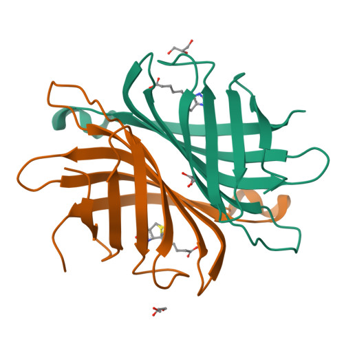
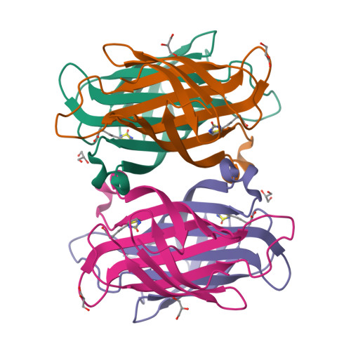
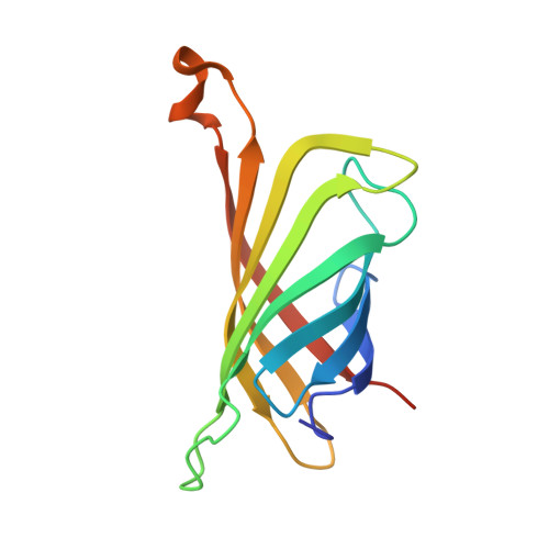
2F01

Epi-biotin complex with core streptavidin


Experimental Data Snapshot

  • Method: X-RAY DIFFRACTION
  • Resolution: 0.85 Å
  • R-Value Free: 0.174 
  • R-Value Work: 0.155 
  • R-Value Observed: 0.156 

Starting Model: experimental
View more details

wwPDB Validation   3D Report Full Report


Ligand Structure Quality Assessment 


This is version 1.3 of the entry. See complete history


Literature

The high-resolution structure of (+)-epi-biotin bound to streptavidin.

Le Trong, I.Aubert, D.G.Thomas, N.R.Stenkamp, R.E.

(2006) Acta Crystallogr D Biol Crystallogr 62: 576-581

  • DOI: https://doi.org/10.1107/S0907444906011887
  • Primary Citation of Related Structures:  
    2F01, 2GH7

  • PubMed Abstract: 

    (+)-Epi-biotin differs from (+)-biotin in the configuration of the chiral center at atom C2. This could lead to a difference in the mode of binding of (+)-epi-biotin to streptavidin, a natural protein receptor for (+)-biotin. Diffraction data were collected to a maximum of 0.85 Angstrom resolution for structural analysis of the complex of streptavidin with a sample of (+)-epi-biotin and refinement was carried out at both 1.0 and 0.85 Angstrom resolution. The structure determination shows a superposition of two ligands in the binding site, (+)-biotin and (+)-epi-biotin. The molecules overlap in the model for the complex except for the position of S1 in the tetrahydrothiophene ring. Differences in the conformation of the ring permits binding of each molecule to streptavidin with little observable difference in the protein structures at this high resolution.


  • Organizational Affiliation

    Departments of Biological Structure and Biochemistry, Biomolecular Structure Center, University of Washington, Seattle, WA 98195, USA.


Macromolecules
Find similar proteins by:  (by identity cutoff)  |  3D Structure
Entity ID: 1
MoleculeChains Sequence LengthOrganismDetailsImage
Streptavidin
A, B
127Streptomyces avidiniiMutation(s): 0 
UniProt
Find proteins for P22629 (Streptomyces avidinii)
Explore P22629 
Go to UniProtKB:  P22629
Entity Groups  
Sequence Clusters30% Identity50% Identity70% Identity90% Identity95% Identity100% Identity
UniProt GroupP22629
Sequence Annotations
Expand
  • Reference Sequence
Small Molecules
Ligands 3 Unique
IDChains Name / Formula / InChI Key2D Diagram3D Interactions
BTQ
Query on BTQ

Download Ideal Coordinates CCD File 
D [auth A],
H [auth B]
EPI-BIOTIN
C10 H16 N2 O3 S
YBJHBAHKTGYVGT-OOZYFLPDSA-N
BTN
Query on BTN

Download Ideal Coordinates CCD File 
C [auth A],
G [auth B]
BIOTIN
C10 H16 N2 O3 S
YBJHBAHKTGYVGT-ZKWXMUAHSA-N
GOL
Query on GOL

Download Ideal Coordinates CCD File 
E [auth A],
F [auth A],
I [auth B]
GLYCEROL
C3 H8 O3
PEDCQBHIVMGVHV-UHFFFAOYSA-N
Binding Affinity Annotations 
IDSourceBinding Affinity
BTN PDBBind:  2F01 Kd: 1.00e-4 (nM) from 1 assay(s)
BindingDB:  2F01 Kd: 0 (nM) from 1 assay(s)
ΔH: min: -1.23e+2, max: -6.69e+1 (kJ/mol) from 12 assay(s)
Experimental Data & Validation

Experimental Data

  • Method: X-RAY DIFFRACTION
  • Resolution: 0.85 Å
  • R-Value Free: 0.174 
  • R-Value Work: 0.155 
  • R-Value Observed: 0.156 
  • Space Group: I 2 2 2
Unit Cell:
Length ( Å )Angle ( ˚ )
a = 46.39α = 90
b = 93.811β = 90
c = 104.449γ = 90
Software Package:
Software NamePurpose
SHELXmodel building
SHELXL-97refinement
DENZOdata reduction
SCALEPACKdata scaling
SHELXphasing

Structure Validation

View Full Validation Report


Ligand Structure Quality Assessment 


Entry History 

Deposition Data

Revision History  (Full details and data files)

  • Version 1.0: 2005-11-29
    Type: Initial release
  • Version 1.1: 2008-05-01
    Changes: Version format compliance
  • Version 1.2: 2011-07-13
    Changes: Non-polymer description, Version format compliance
  • Version 1.3: 2023-08-23
    Changes: Data collection, Database references, Derived calculations, Refinement description