Nucleotide Substrate Recognition by Udp-N-Acetylglucosamine Acyltransferase (Lpxa) in the First Step of Lipid a Biosynthesis.
Ulaganathan, V., Buetow, L., Hunter, W.N.(2007) J Mol Biology 369: 305
- PubMed: 17434525
- DOI: https://doi.org/10.1016/j.jmb.2007.03.039
- Primary Citation of Related Structures:
2JF2, 2JF3 - PubMed Abstract:
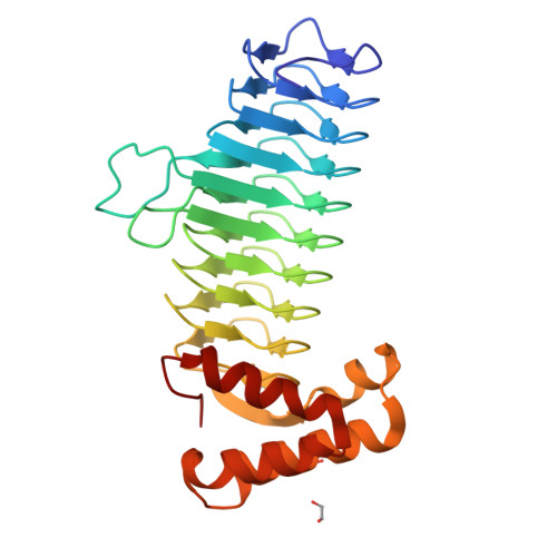
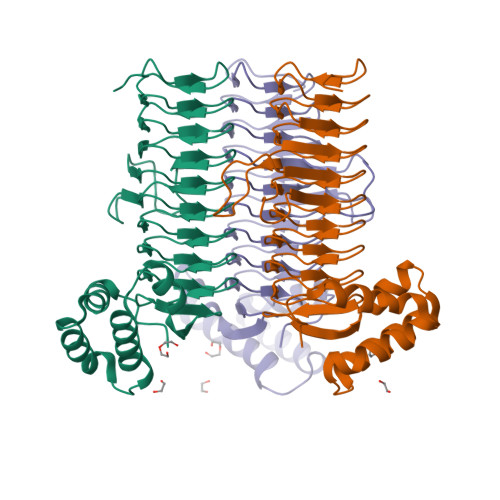
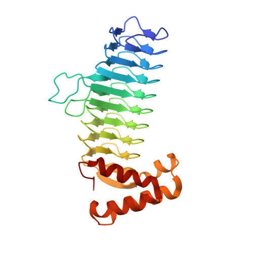
Lipid A is an integral component of the lipopolysaccharide (LPS) that forms the selective and protective outer monolayer of Gram-negative bacteria, and is essential for bacterial growth and viability. UDP-N-acetylglucosamine acyltransferase (LpxA) initiates lipid A biosynthesis by catalyzing the transfer of R-3-hydroxymyristic acid from acyl carrier protein to the 3'-hydroxyl group of UDP-GlcNAc. The enzyme is a homotrimer, and previous studies suggested that the active site lies within a positively charged cleft formed at the subunit-subunit interface. The crystal structure of Escherichia coli LpxA in complex with UDP-GlcNAc reveals details of the substrate-binding site, with prominent hydrophilic interactions between highly conserved clusters of residues (Asn198, Glu200, Arg204 and Arg205) with UDP, and (Asp74, His125, His144 and Gln161) with the GlcNAc moiety. These interactions serve to bind and orient the substrate for catalysis. The crystallographic model supports previous results, which suggest that acylation occurs via nucleophilic attack of deprotonated UDP-GlcNAc on the acyl donor in a general base-catalyzed mechanism involving a catalytic dyad of His125 and Asp126. His125, the general base, interacts with the 3'-hydroxyl group of UDP-GlcNAc to generate the nucleophile. The Asp126 side-chain accepts a hydrogen bond from His125 and helps orient the general base to participate in catalysis. Comparisons with an LpxA:peptide inhibitor complex indicate that the peptide competes with both nucleotide and acyl carrier protein substrates.
Organizational Affiliation:
Division of Biological Chemistry and Molecular Microbiology, College of Life Sciences, University of Dundee, Dundee DD1 5EH, UK.


