2V95 | pdb_00002v95

Structure of Corticosteroid-Binding Globulin in complex with Cortisol


Experimental Data Snapshot

  • Method: X-RAY DIFFRACTION
  • Resolution: 1.93 Å
  • R-Value Free: 
    0.264 (Depositor), 0.270 (DCC) 
  • R-Value Work: 
    0.210 (Depositor), 0.210 (DCC) 
  • R-Value Observed: 
    0.214 (Depositor) 

Starting Model: experimental
View more details

wwPDB Validation   3D Report Full Report


Ligand Structure Quality Assessment 

Created with Raphaël 2.3.0Worse 01 BetterLigand structure goodness of fit to experimental dataBest fitted HCYClick on this verticalbar to view details

This is version 1.2 of the entry. See complete history


Literature

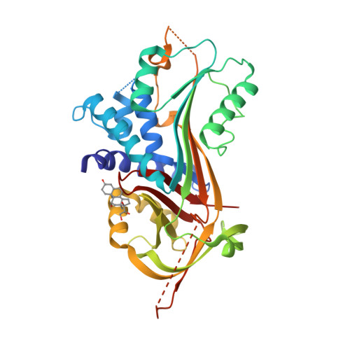
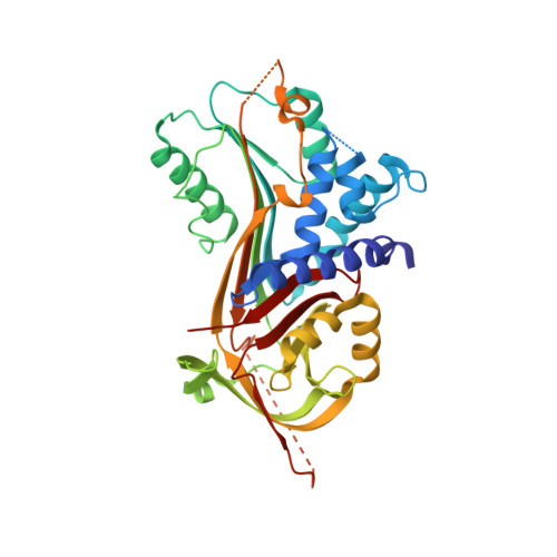
Corticosteroid-Binding Globulin: Structural Basis for Steroid Transport and Proteinase-Triggered Release

Klieber, M.A.Underhill, C.Hammond, G.L.Muller, Y.A.

(2007) J Biological Chem 282: 29594

  • DOI: https://doi.org/10.1074/jbc.M705014200
  • Primary Citation of Related Structures:  
    2V95

  • PubMed Abstract: 

    Corticosteroid-binding globulin (CBG) is a serine proteinase inhibitor (serpin) family member that transports glucocorticoids in blood and regulates their access to target cells. The 1.9A crystal structure of rat CBG shows that its steroid-binding site resembles the thyroxin-binding site in the related serpin, thyroxin-binding globulin, and mutagenesis studies have confirmed the contributions of key residues that constitute the steroid-binding pocket. Unlike thyroxin-bound thyroxin-binding globulin, the cortisol-bound CBG displays an "active" serpin conformation with the proteinase-sensitive, reactive center loop (RCL) fully expelled from the regulatory beta-sheet A. Moreover, the CBG structure allows us to predict that complete insertion of the proteolytically cleaved RCL into the serpin fold occurs in concert with a displacement and unwinding of helix D that would disrupt the steroid-binding site. This allosteric coupling between RCL positioning and occupancy of the CBG steroid-binding site, which resembles the ligand (glycosamino-glycan)-dependent activation of the thrombin inhibitory serpins heparin cofactor II and anti-thrombin RCLs, ensures both optimal recognition of CBG by target proteinases and efficient release of steroid to sites of action.


  • Organizational Affiliation

    Lehrstuhl für Biotechnik, Department of Biology, Friedrich-Alexander-University Erlangen-Nuremberg, D-91052 Erlangen, Germany.


Macromolecules
Find similar proteins by:  (by identity cutoff)  |  3D Structure
Entity ID: 1
MoleculeChains Sequence LengthOrganismDetailsImage
CORTICOSTEROID-BINDING GLOBULIN371Rattus norvegicusMutation(s): 2 
UniProt
Find proteins for P31211 (Rattus norvegicus)
Explore P31211 
Go to UniProtKB:  P31211
Entity Groups  
Sequence Clusters30% Identity50% Identity70% Identity90% Identity95% Identity100% Identity
UniProt GroupP31211
Sequence Annotations
Expand
  • Reference Sequence
Small Molecules
Ligands 1 Unique
IDChains Name / Formula / InChI Key2D Diagram3D Interactions
HCY
Query on HCY

Download Ideal Coordinates CCD File 
B [auth A](11alpha,14beta)-11,17,21-trihydroxypregn-4-ene-3,20-dione
C21 H30 O5
JYGXADMDTFJGBT-VWUMJDOOSA-N
Experimental Data & Validation

Experimental Data

  • Method: X-RAY DIFFRACTION
  • Resolution: 1.93 Å
  • R-Value Free:  0.264 (Depositor), 0.270 (DCC) 
  • R-Value Work:  0.210 (Depositor), 0.210 (DCC) 
  • R-Value Observed: 0.214 (Depositor) 
Space Group: C 1 2 1
Unit Cell:
Length ( Å )Angle ( ˚ )
a = 122.11α = 90
b = 54.25β = 97.16
c = 61.04γ = 90
Software Package:
Software NamePurpose
REFMACrefinement
XDSdata reduction
XSCALEdata scaling
AMoREphasing

Structure Validation

View Full Validation Report


Ligand Structure Quality Assessment 

Created with Raphaël 2.3.0Worse 01 BetterLigand structure goodness of fit to experimental dataBest fitted HCYClick on this verticalbar to view details

Entry History 

Deposition Data

Revision History  (Full details and data files)

  • Version 1.0: 2007-09-04
    Type: Initial release
  • Version 1.1: 2015-12-02
    Changes: Derived calculations, Other, Refinement description, Structure summary, Version format compliance
  • Version 1.2: 2023-12-13
    Changes: Data collection, Database references, Derived calculations, Other, Refinement description