Fucose-Derived Glycomimetics as High Affinity Ligands for Bacterial Lectin Pa-Iil from Pseudomonas Aeruginosa
Beha, S., Marotte, K., Sabin, C., Mitchell, E.P., Imberty, A., Roy, R.To be published.
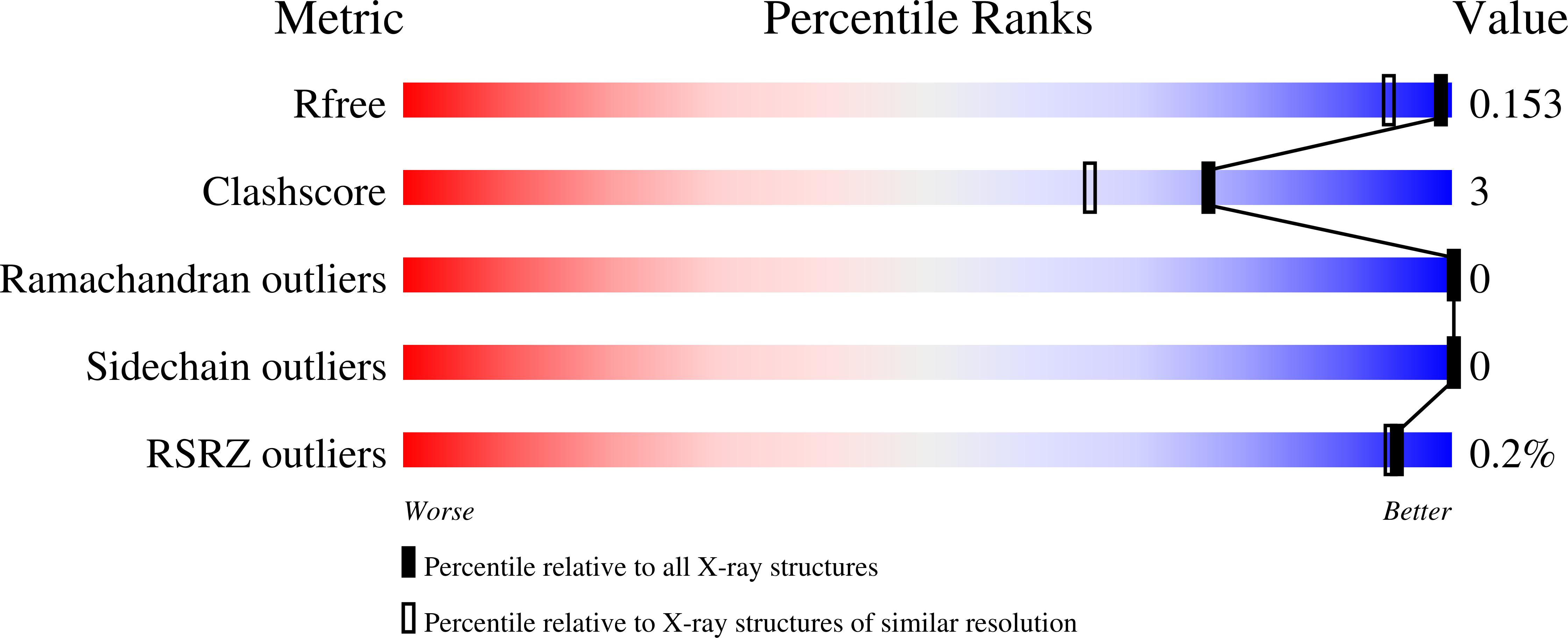
Experimental Data Snapshot
Starting Model: experimental
View more details
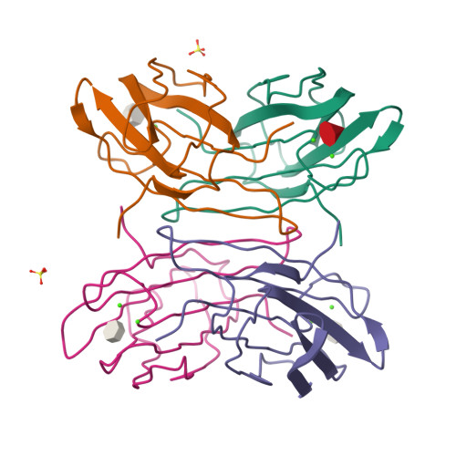
Entity ID: 1 | |||||
|---|---|---|---|---|---|
| Molecule | Chains | Sequence Length | Organism | Details | Image |
| FUCOSE-BINDING LECTIN PA-IIL | 114 | Pseudomonas aeruginosa | Mutation(s): 0 | 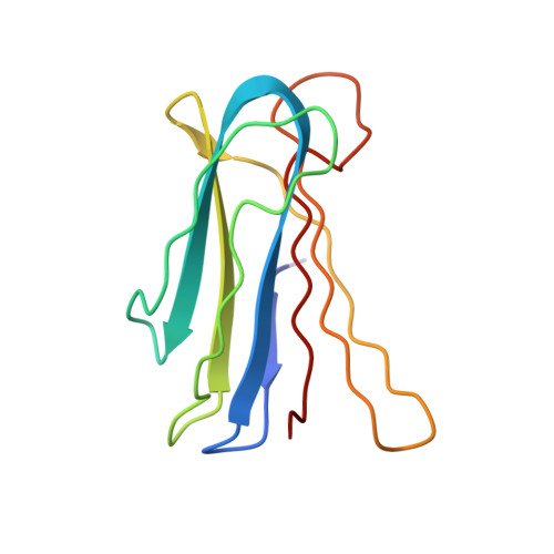 | |
UniProt | |||||
Find proteins for Q9HYN5 (Pseudomonas aeruginosa (strain ATCC 15692 / DSM 22644 / CIP 104116 / JCM 14847 / LMG 12228 / 1C / PRS 101 / PAO1)) Explore Q9HYN5 Go to UniProtKB: Q9HYN5 | |||||
Entity Groups | |||||
| Sequence Clusters | 30% Identity50% Identity70% Identity90% Identity95% Identity100% Identity | ||||
| UniProt Group | Q9HYN5 | ||||
Sequence AnnotationsExpand | |||||
| |||||
| Ligands 4 Unique | |||||
|---|---|---|---|---|---|
| ID | Chains | Name / Formula / InChI Key | 2D Diagram | 3D Interactions | |
| YX0 Query on YX0 | I [auth B], M [auth C], P [auth D] | [(3E)-3-(1-hydroxyethylidene)-2,3-dihydroisoxazol-5-yl]methyl alpha-L-fucopyranoside C12 H19 N O7 DQRDRRBASDQFKR-AWJQDWPASA-N |  | ||
| FUC Query on FUC | E [auth A] | alpha-L-fucopyranose C6 H12 O5 SHZGCJCMOBCMKK-SXUWKVJYSA-N |  | ||
| SO4 Query on SO4 | H [auth A], J [auth B] | SULFATE ION O4 S QAOWNCQODCNURD-UHFFFAOYSA-L |  | ||
| CA Query on CA | F [auth A] G [auth A] K [auth B] L [auth B] N [auth C] | CALCIUM ION Ca BHPQYMZQTOCNFJ-UHFFFAOYSA-N |  | ||
| Length ( Å ) | Angle ( ˚ ) |
|---|---|
| a = 52.555 | α = 90 |
| b = 72.668 | β = 94.42 |
| c = 54.725 | γ = 90 |
| Software Name | Purpose |
|---|---|
| REFMAC | refinement |
| MOSFLM | data reduction |
| SCALA | data scaling |
| MOLREP | phasing |