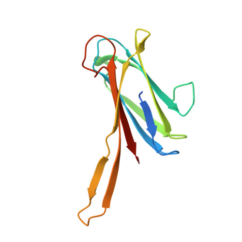
2WR9

CRYSTAL STRUCTURE OF BURKHOLDERIA CENOCEPACIA LECTIN (BCLA) COMPLEXED WITH AMAN1-3MAN DISACCHARIDE


Experimental Data Snapshot

  • Method: X-RAY DIFFRACTION
  • Resolution: 1.75 Å
  • R-Value Free: 0.225 
  • R-Value Work: 0.176 
  • R-Value Observed: 0.179 

Starting Model: experimental
View more details

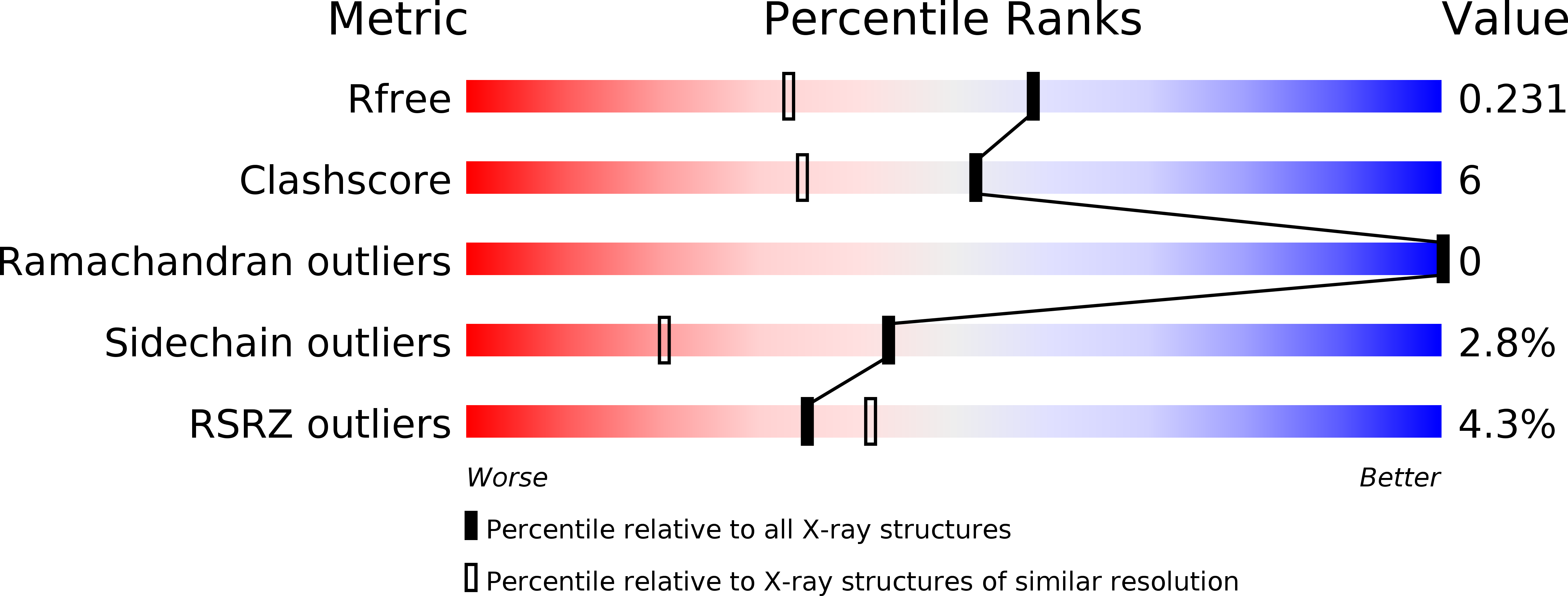
wwPDB Validation   3D Report Full Report


Ligand Structure Quality Assessment 


This is version 2.1 of the entry. See complete history


Literature

Structural Basis of the Affinity for Oligomannosides and Analogs Displayed by Bc2L-A, a Burkholderia Cenocepacia Soluble Lectin.

Lameignere, E.Shiao, T.C.Roy, R.Wimmerova, M.Dubreuil, F.Varrot, A.Imberty, A.

(2010) Glycobiology 20: 87

  • DOI: https://doi.org/10.1093/glycob/cwp151
  • Primary Citation of Related Structures:  
    2WR9, 2WRA

  • PubMed Abstract: 

    The opportunistic pathogen Burkholderia cenocepacia contains three soluble carbohydrate-binding proteins, related to the fucose-binding lectin PA-IIL from Pseudomonas aeruginosa. All contain a PA-IIL-like domain and two of them have an additional N-terminal domain that displays no sequence similarities with known proteins. Printed arrays screening performed on the shortest one, B. cenocepacia lectin A (BC2L-A), demonstrated the strict specificity for oligomannose-type N-glycan structures (Lameignere E, Malinovská L, Sláviková M, Duchaud E, Mitchell EP, Varrot A, Sedo O, Imberty A, Wimmerová M. 2008. Structural basis for mannose recognition by a lectin from opportunistic bacteria Burkholderia cenocepacia. Biochem J. 411:307-318.). The disaccharides alphaMan1-2Man, alphaMan1-3Man, and alphaMan1-6Man and the trisaccharide alphaMan1-3(alphaMan1-6)Man were tested by titration microcalorimetry in order to evaluate their affinity for BC2L-A in solution and to characterize the thermodynamics of the binding. Oligomannose analogs presenting two mannoside residues separated by either flexible or rigid spacer were also tested. Only the rigid one yields to high affinity binding with a fast kinetics of clustering, while the flexible analog and the trimannoside display moderate affinities and no clustering effect on short time scale. The crystal structures of BC2L-A have been obtained in complex with alphaMan1-3Man disaccharide and alphaMan1(alphaMan1-6)-3Man trisaccharide. The lengthy time required for the co-crystallization with the trisaccharide allowed for the formation of cluster since in the BC2L-A-trimannose complex solved at 1.1 A resolution, the sugar creates a bridge between two adjacent dimers, yielding to molecular strings. AFM experiments were performed in order to visualize the filaments formed in solution by this type of interaction.


  • Organizational Affiliation

    CERMAV-CNRS (affiliated with Université Joseph Fourier and belonging to ICMG), BP 53, F-38041, Grenoble, Cedex 09, France.


Macromolecules
Find similar proteins by:  (by identity cutoff)  |  3D Structure
Entity ID: 1
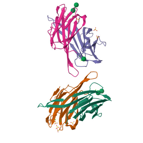
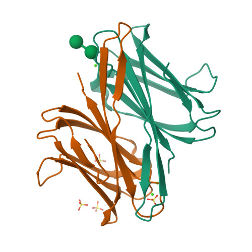
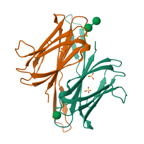
MoleculeChains Sequence LengthOrganismDetailsImage
LECTIN
A, B, C, D
128Burkholderia cenocepacia J2315Mutation(s): 0 
UniProt
Find proteins for B4EH87 (Burkholderia cenocepacia (strain ATCC BAA-245 / DSM 16553 / LMG 16656 / NCTC 13227 / J2315 / CF5610))
Explore B4EH87 
Go to UniProtKB:  B4EH87
Entity Groups  
Sequence Clusters30% Identity50% Identity70% Identity90% Identity95% Identity100% Identity
UniProt GroupB4EH87
Sequence Annotations
Expand
  • Reference Sequence
Oligosaccharides

Help

Entity ID: 2
MoleculeChains Length2D Diagram Glycosylation3D Interactions
alpha-D-mannopyranose-(1-3)-alpha-D-mannopyranose
E, F
2N/A
Glycosylation Resources
GlyTouCan:  G00891MP
GlyCosmos:  G00891MP
GlyGen:  G00891MP
Small Molecules
Ligands 3 Unique
IDChains Name / Formula / InChI Key2D Diagram3D Interactions
MAN
Query on MAN

Download Ideal Coordinates CCD File 
O [auth C]alpha-D-mannopyranose
C6 H12 O6
WQZGKKKJIJFFOK-PQMKYFCFSA-N
SO4
Query on SO4

Download Ideal Coordinates CCD File 
K [auth B]
L [auth B]
M [auth B]
N [auth B]
R [auth C]
K [auth B],
L [auth B],
M [auth B],
N [auth B],
R [auth C],
S [auth C],
V [auth D]
SULFATE ION
O4 S
QAOWNCQODCNURD-UHFFFAOYSA-L
CA
Query on CA

Download Ideal Coordinates CCD File 
G [auth A]
H [auth B]
I [auth B]
J [auth B]
P [auth C]
G [auth A],
H [auth B],
I [auth B],
J [auth B],
P [auth C],
Q [auth C],
T [auth D],
U [auth D]
CALCIUM ION
Ca
BHPQYMZQTOCNFJ-UHFFFAOYSA-N
Biologically Interesting Molecules (External Reference) 1 Unique
Experimental Data & Validation

Experimental Data

  • Method: X-RAY DIFFRACTION
  • Resolution: 1.75 Å
  • R-Value Free: 0.225 
  • R-Value Work: 0.176 
  • R-Value Observed: 0.179 
  • Space Group: P 1 21 1
Unit Cell:
Length ( Å )Angle ( ˚ )
a = 57.958α = 90
b = 48.974β = 98.89
c = 76.261γ = 90
Software Package:
Software NamePurpose
REFMACrefinement
MOSFLMdata reduction
SCALAdata scaling
MOLREPphasing

Structure Validation

View Full Validation Report


Ligand Structure Quality Assessment 


Entry History 

Deposition Data

Revision History  (Full details and data files)

  • Version 1.0: 2009-09-15
    Type: Initial release
  • Version 1.1: 2011-08-03
    Changes: Database references, Derived calculations, Non-polymer description, Other, Refinement description, Source and taxonomy, Structure summary, Version format compliance
  • Version 2.0: 2020-07-29
    Type: Remediation
    Reason: Carbohydrate remediation
    Changes: Atomic model, Data collection, Derived calculations, Other, Structure summary
  • Version 2.1: 2023-12-20
    Changes: Data collection, Database references, Derived calculations, Refinement description, Structure summary