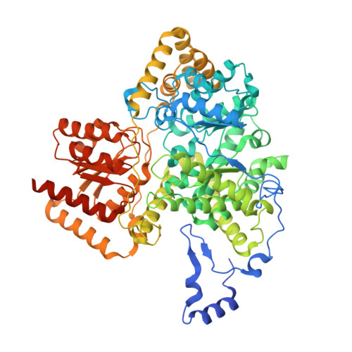
2XIJ

Crystal structure of human methylmalonyl-CoA mutase in complex with adenosylcobalamin


Experimental Data Snapshot

  • Method: X-RAY DIFFRACTION
  • Resolution: 1.95 Å
  • R-Value Free: 0.199 
  • R-Value Work: 0.158 
  • R-Value Observed: 0.160 

Starting Model: experimental
View more details

wwPDB Validation   3D Report Full Report


Ligand Structure Quality Assessment 


This is version 1.2 of the entry. See complete history


Literature

Structures of the Human Gtpase Mmaa and Vitamin B12-Dependent Methylmalonyl-Coa Mutase and Insight Into Their Complex Formation.

Froese, D.S.Kochan, G.Muniz, J.Wu, X.Gileadi, C.Ugochukwu, E.Krysztofinska, E.Gravel, R.A.Oppermann, U.Yue, W.W.

(2010) J Biol Chem 285: 38204

  • DOI: https://doi.org/10.1074/jbc.M110.177717
  • Primary Citation of Related Structures:  
    2WWW, 2XIJ, 2XIQ, 3BIC

  • PubMed Abstract: 

    Vitamin B(12) (cobalamin, Cbl) is essential to the function of two human enzymes, methionine synthase (MS) and methylmalonyl-CoA mutase (MUT). The conversion of dietary Cbl to its cofactor forms, methyl-Cbl (MeCbl) for MS and adenosyl-Cbl (AdoCbl) for MUT, located in the cytosol and mitochondria, respectively, requires a complex pathway of intracellular processing and trafficking. One of the processing proteins, MMAA (methylmalonic aciduria type A), is implicated in the mitochondrial assembly of AdoCbl into MUT and is defective in children from the cblA complementation group of cobalamin disorders. To characterize the functional interplay between MMAA and MUT, we have crystallized human MMAA in the GDP-bound form and human MUT in the apo, holo, and substrate-bound ternary forms. Structures of both proteins reveal highly conserved domain architecture and catalytic machinery for ligand binding, yet they show substantially different dimeric assembly and interaction, compared with their bacterial counterparts. We show that MMAA exhibits GTPase activity that is modulated by MUT and that the two proteins interact in vitro and in vivo. Formation of a stable MMAA-MUT complex is nucleotide-selective for MMAA (GMPPNP over GDP) and apoenzyme-dependent for MUT. The physiological importance of this interaction is highlighted by a recently identified homoallelic patient mutation of MMAA, G188R, which, we show, retains basal GTPase activity but has abrogated interaction. Together, our data point to a gatekeeping role for MMAA by favoring complex formation with MUT apoenzyme for AdoCbl assembly and releasing the AdoCbl-loaded holoenzyme from the complex, in a GTP-dependent manner.


  • Organizational Affiliation

    Structural Genomics Consortium, University of Oxford OX3 7DU, United Kingdom.


Macromolecules
Find similar proteins by:  (by identity cutoff)  |  3D Structure
Entity ID: 1
MoleculeChains Sequence LengthOrganismDetailsImage
METHYLMALONYL-COA MUTASE, MITOCHONDRIAL762Homo sapiensMutation(s): 1 
EC: 5.4.99.2
UniProt & NIH Common Fund Data Resources
Find proteins for P22033 (Homo sapiens)
Explore P22033 
Go to UniProtKB:  P22033
PHAROS:  P22033
GTEx:  ENSG00000146085 
Entity Groups  
Sequence Clusters30% Identity50% Identity70% Identity90% Identity95% Identity100% Identity
UniProt GroupP22033
Sequence Annotations
Expand
  • Reference Sequence
Small Molecules
Ligands 5 Unique
IDChains Name / Formula / InChI Key2D Diagram3D Interactions
B12
Query on B12

Download Ideal Coordinates CCD File 
N [auth A]COBALAMIN
C62 H89 Co N13 O14 P
LKVIQTCSMMVGFU-DWSMJLPVSA-N
5AD
Query on 5AD

Download Ideal Coordinates CCD File 
B [auth A]5'-DEOXYADENOSINE
C10 H13 N5 O3
XGYIMTFOTBMPFP-KQYNXXCUSA-N
BTB
Query on BTB

Download Ideal Coordinates CCD File 
L [auth A]2-[BIS-(2-HYDROXY-ETHYL)-AMINO]-2-HYDROXYMETHYL-PROPANE-1,3-DIOL
C8 H19 N O5
OWMVSZAMULFTJU-UHFFFAOYSA-N
SO4
Query on SO4

Download Ideal Coordinates CCD File 
C [auth A],
D [auth A]
SULFATE ION
O4 S
QAOWNCQODCNURD-UHFFFAOYSA-L
EDO
Query on EDO

Download Ideal Coordinates CCD File 
E [auth A]
F [auth A]
G [auth A]
H [auth A]
I [auth A]
E [auth A],
F [auth A],
G [auth A],
H [auth A],
I [auth A],
J [auth A],
K [auth A],
M [auth A]
1,2-ETHANEDIOL
C2 H6 O2
LYCAIKOWRPUZTN-UHFFFAOYSA-N
Experimental Data & Validation

Experimental Data

  • Method: X-RAY DIFFRACTION
  • Resolution: 1.95 Å
  • R-Value Free: 0.199 
  • R-Value Work: 0.158 
  • R-Value Observed: 0.160 
  • Space Group: C 2 2 21
Unit Cell:
Length ( Å )Angle ( ˚ )
a = 59.36α = 90
b = 137.72β = 90
c = 198.4γ = 90
Software Package:
Software NamePurpose
REFMACrefinement
MOSFLMdata reduction
SCALAdata scaling
PHASERphasing

Structure Validation

View Full Validation Report


Ligand Structure Quality Assessment 


Entry History 

Revision History  (Full details and data files)

  • Version 1.0: 2010-09-29
    Type: Initial release
  • Version 1.1: 2012-10-31
    Changes: Atomic model, Derived calculations, Non-polymer description, Other, Version format compliance
  • Version 1.2: 2023-12-20
    Changes: Data collection, Database references, Derived calculations, Other, Refinement description