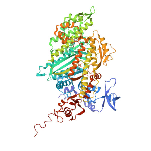
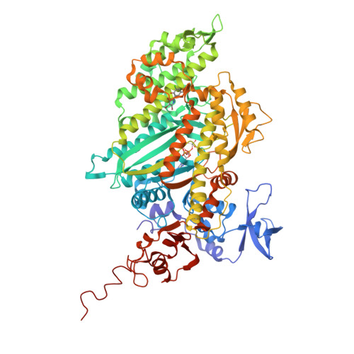
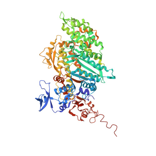
2XO8

Crystal Structure of Myosin-2 in Complex with Tribromodichloropseudilin


Experimental Data Snapshot

  • Method: X-RAY DIFFRACTION
  • Resolution: 2.40 Å
  • R-Value Free: 0.254 
  • R-Value Work: 0.202 
  • R-Value Observed: 0.202 

Starting Model: experimental
View more details

wwPDB Validation   3D Report Full Report


Ligand Structure Quality Assessment 


This is version 1.5 of the entry. See complete history


Literature

Inhibition of Myosin ATPase Activity by Halogenated Pseudilins: A Structure-Activity Study.

Preller, M.Chinthalapudi, K.Martin, R.Knolker, H.Manstein, D.J.

(2011) J Med Chem 54: 3675

  • DOI: https://doi.org/10.1021/jm200259f
  • Primary Citation of Related Structures:  
    2XO8

  • PubMed Abstract: 

    Myosin activity is crucial for many biological functions. Strong links have been established between changes in the activity of specific myosin isoforms and diseases such as cancer, cardiovascular failure, and disorders of sensory organs and the central nervous system. The modulation of specific myosin isoforms therefore holds a strong therapeutic potential. In recent work, we identified members of the marine alkaloid family of pseudilins as potent inhibitors of myosin-dependent processes. Here, we report the crystal structure of the complex between the Dictyostelium myosin 2 motor domain and 2,4-dichloro-6-(3,4,5-tribromo-1H-pyrrole-2-yl)phenol (3). Detailed comparison with previously solved structures of the myosin 2 complex with bound pentabromopseudilin (2a) or pentachloropseudilin (4a) provides insights into the molecular basis of the allosteric communication between the catalytic and the allosteric sites. Moreover, we describe the inhibitory potency for a congeneric series of halogenated pseudilins. Insight into their mode of action is gained by applying a combination of experimental and computational approaches.


  • Organizational Affiliation

    Institute for Biophysical Chemistry, Hannover Medical School, 30625 Hannover,


Macromolecules
Find similar proteins by:  (by identity cutoff)  |  3D Structure
Entity ID: 1
MoleculeChains Sequence LengthOrganismDetailsImage
MYOSIN-2 HEAVY CHAIN776Dictyostelium discoideumMutation(s): 0 
UniProt
Find proteins for P08799 (Dictyostelium discoideum)
Explore P08799 
Go to UniProtKB:  P08799
Entity Groups  
Sequence Clusters30% Identity50% Identity70% Identity90% Identity95% Identity100% Identity
UniProt GroupP08799
Sequence Annotations
Expand
  • Reference Sequence
Small Molecules
Binding Affinity Annotations 
IDSourceBinding Affinity
H70 PDBBind:  2XO8 IC50: 4.72e+4 (nM) from 1 assay(s)
Experimental Data & Validation

Experimental Data

  • Method: X-RAY DIFFRACTION
  • Resolution: 2.40 Å
  • R-Value Free: 0.254 
  • R-Value Work: 0.202 
  • R-Value Observed: 0.202 
  • Space Group: C 2 2 21
Unit Cell:
Length ( Å )Angle ( ˚ )
a = 88.34α = 90
b = 146.383β = 90
c = 152.798γ = 90
Software Package:
Software NamePurpose
CNSrefinement
XDSdata reduction
XDSdata scaling
CNSphasing

Structure Validation

View Full Validation Report


Ligand Structure Quality Assessment 


Entry History 

Deposition Data

Revision History  (Full details and data files)

  • Version 1.0: 2011-05-25
    Type: Initial release
  • Version 1.1: 2013-01-30
    Changes: Database references, Version format compliance
  • Version 1.2: 2018-06-20
    Changes: Advisory, Data collection, Derived calculations
  • Version 1.3: 2019-07-10
    Changes: Data collection
  • Version 1.4: 2019-07-24
    Changes: Data collection
  • Version 1.5: 2023-12-20
    Changes: Advisory, Data collection, Database references, Derived calculations, Other, Refinement description