2ZA1

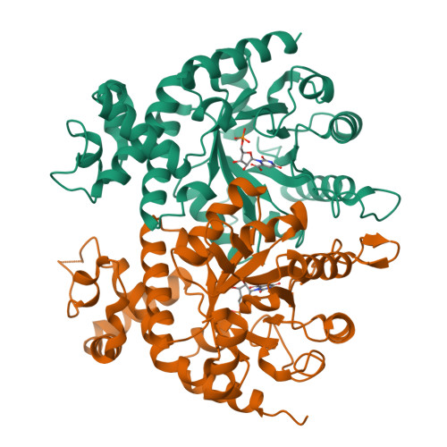
Crystal Structure of orotidine 5'-monophosphate decarboxylase complexed with orotidine 5'-monophosphate from P.falciparum


Experimental Data Snapshot

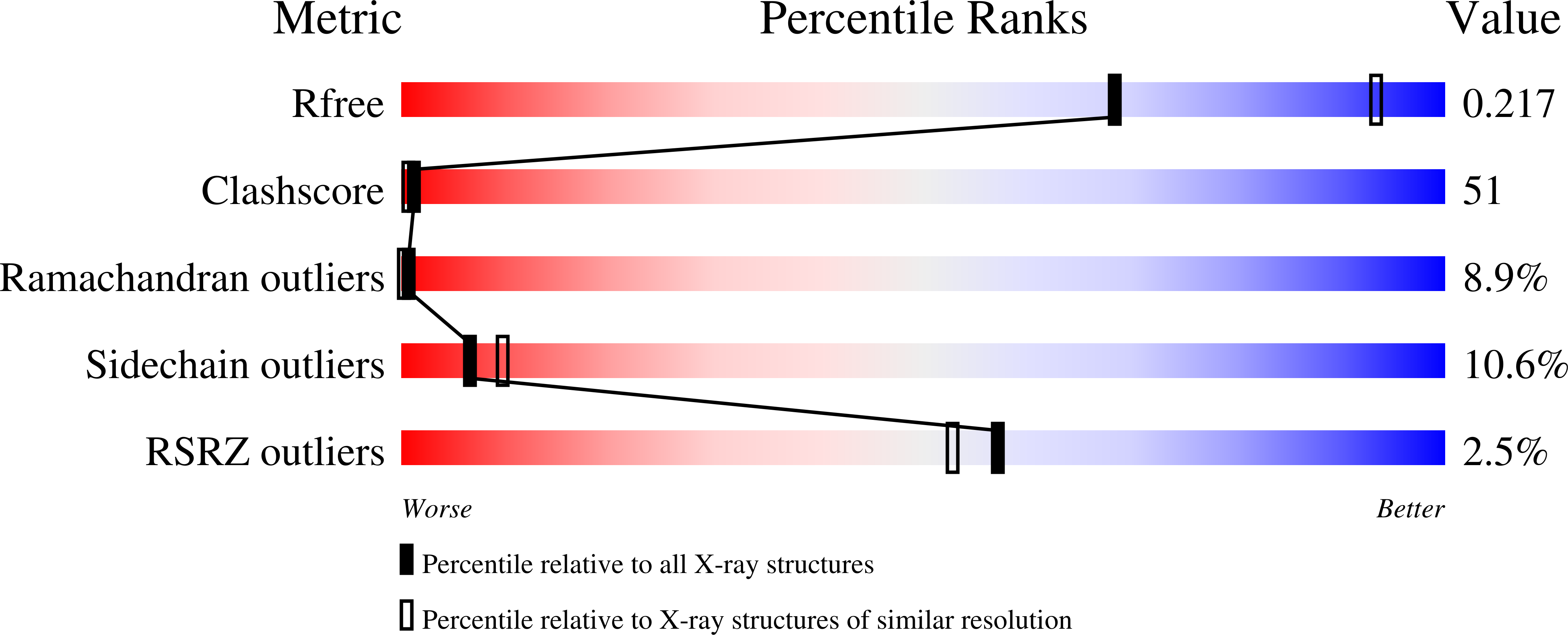
  • Method: X-RAY DIFFRACTION
  • Resolution: 2.65 Å
  • R-Value Free: 0.292 
  • R-Value Work: 0.210 
  • R-Value Observed: 0.210 

Starting Model: experimental
View more details

wwPDB Validation   3D Report Full Report


Ligand Structure Quality Assessment 


This is version 1.2 of the entry. See complete history

Re-refinement Note

A newer entry is available that reflects an alternative modeling of the original data: 6DSR


Literature

Structural Basis for the Decarboxylation of Orotidine 5'-Monophosphate (OMP) by Plasmodium Falciparum OMP Decarboxylase

Tokuoka, K.Kusakari, Y.Krungkrai, S.R.Matsumura, H.Kai, Y.Krungkrai, J.Horii, T.Inoue, T.

(2008) J Biochem 143: 69-78

  • DOI: https://doi.org/10.1093/jb/mvm193
  • Primary Citation of Related Structures:  
    2ZA1, 2ZA2, 2ZA3

  • PubMed Abstract: 

    Orotidine 5'-monophoshate decarboxylase (OMPDC) catalyses the decarboxylation of orotidine 5'-monophosphate (OMP) to uridine 5'-monophosphate (UMP). Here, we report the X-ray analysis of apo, substrate or product-complex forms of OMPDC from Plasmodium falciparum (PfOMPDC) at 2.7, 2.65 and 2.65 A, respectively. The structural analysis provides the substrate recognition mechanism with dynamic structural changes, as well as the rearrangement of the hydrogen bond array at the active site. The structural basis of substrate or product binding to PfOMPDC will help to uncover the decarboxylation mechanism and facilitate structure-based optimization of antimalarial drugs.


  • Organizational Affiliation

    Department of Applied Chemistry, Graduate School of Engineering, Osaka University, 2-1 Yamadaoka, Suita, Osaka 565-0871, Japan.


Macromolecules
Find similar proteins by:  (by identity cutoff)  |  3D Structure
Entity ID: 1
MoleculeChains Sequence LengthOrganismDetailsImage
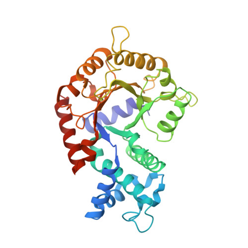
Orotidine 5'-phosphate decarboxylase
A, B
323Plasmodium falciparumMutation(s): 0 
EC: 4.1.1.23
UniProt
Find proteins for Q8T6J6 (Plasmodium falciparum)
Explore Q8T6J6 
Go to UniProtKB:  Q8T6J6
Entity Groups  
Sequence Clusters30% Identity50% Identity70% Identity90% Identity95% Identity100% Identity
UniProt GroupQ8T6J6
Sequence Annotations
Expand
  • Reference Sequence
Experimental Data & Validation

Experimental Data

  • Method: X-RAY DIFFRACTION
  • Resolution: 2.65 Å
  • R-Value Free: 0.292 
  • R-Value Work: 0.210 
  • R-Value Observed: 0.210 
  • Space Group: H 3
Unit Cell:
Length ( Å )Angle ( ˚ )
a = 201.8α = 90
b = 201.8β = 90
c = 44.2γ = 120
Software Package:
Software NamePurpose
CNSrefinement
HKL-2000data reduction
HKL-2000data scaling
AMoREphasing

Structure Validation

View Full Validation Report


Ligand Structure Quality Assessment 


Entry History 

Deposition Data

Revision History  (Full details and data files)

  • Version 1.0: 2007-12-04
    Type: Initial release
  • Version 1.1: 2011-07-13
    Changes: Version format compliance
  • Version 1.2: 2023-11-01
    Changes: Data collection, Database references, Derived calculations, Refinement description