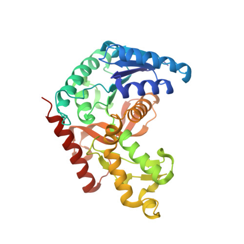
2AA3

Crystal structure of Plasmodium vivax lactate dehydrogenase complex with APADH


Experimental Data Snapshot

  • Method: X-RAY DIFFRACTION
  • Resolution: 2.05 Å
  • R-Value Free: 0.226 
  • R-Value Work: 0.191 
  • R-Value Observed: 0.192 

Starting Model: experimental
View more details

wwPDB Validation   3D Report Full Report


Ligand Structure Quality Assessment 


This is version 2.0 of the entry. See complete history


Literature

Structure of Lactate Dehydrogenase from Plasmodium vivax: Complexes with NADH and APADH.

Chaikuad, A.Fairweather, V.Conners, R.Joseph-Horne, T.Turgut-Balik, D.Brady, R.L.

(2005) Biochemistry 44: 16221-16228

  • DOI: https://doi.org/10.1021/bi051416y
  • Primary Citation of Related Structures:  
    2A92, 2A94, 2AA3

  • PubMed Abstract: 

    Malaria caused by Plasmodium vivax is a major cause of global morbidity and, in rare cases, mortality. Lactate dehydrogenase is an essential Plasmodium protein and, therefore, a potential antimalarial drug target. Ideally, drugs directed against this target would be effective against both major species of Plasmodium, P. falciparum and P. vivax. In this study, the crystal structure of the lactate dehydrogenase protein from P. vivax has been solved and is compared to the equivalent structure from the P. falciparum enzyme. The active sites and cofactor binding pockets of both enzymes are found to be highly similar and differentiate these enzymes from their human counterparts. These structures suggest effective inhibition of both enzymes should be readily achievable with a common inhibitor. The crystal structures of both enzymes have also been solved in complex with the synthetic cofactor APADH. The unusual cofactor binding site in these Plasmodium enzymes is found to readily accommodate both NADH and APADH, explaining why the Plasmodium enzymes retain enzymatic activity in the presence of this synthetic cofactor.


  • Organizational Affiliation

    Department of Biochemistry, University of Bristol, United Kingdom.


Macromolecules
Find similar proteins by:  (by identity cutoff)  |  3D Structure
Entity ID: 1
MoleculeChains Sequence LengthOrganismDetailsImage
L-lactate dehydrogenase
A, B, C, D
321Plasmodium vivaxMutation(s): 0 
Gene Names: LDH
EC: 1.1.1.27
UniProt
Find proteins for Q4PRK9 (Plasmodium vivax)
Explore Q4PRK9 
Go to UniProtKB:  Q4PRK9
Entity Groups  
Sequence Clusters30% Identity50% Identity70% Identity90% Identity95% Identity100% Identity
UniProt GroupQ4PRK9
Sequence Annotations
Expand
  • Reference Sequence
Experimental Data & Validation

Experimental Data

  • Method: X-RAY DIFFRACTION
  • Resolution: 2.05 Å
  • R-Value Free: 0.226 
  • R-Value Work: 0.191 
  • R-Value Observed: 0.192 
  • Space Group: P 21 21 21
Unit Cell:
Length ( Å )Angle ( ˚ )
a = 81.482α = 90
b = 128.454β = 90
c = 130.781γ = 90
Software Package:
Software NamePurpose
DENZOdata reduction
SCALEPACKdata scaling
AMoREphasing
REFMACrefinement
PDB_EXTRACTdata extraction
HKL-2000data reduction

Structure Validation

View Full Validation Report


Ligand Structure Quality Assessment 


Entry History 

Deposition Data

Revision History  (Full details and data files)

  • Version 1.0: 2006-01-10
    Type: Initial release
  • Version 1.1: 2008-04-30
    Changes: Version format compliance
  • Version 1.2: 2011-07-13
    Changes: Version format compliance
  • Version 1.3: 2017-10-11
    Changes: Advisory, Refinement description
  • Version 1.4: 2023-08-23
    Changes: Advisory, Data collection, Database references, Derived calculations, Refinement description
  • Version 2.0: 2023-09-20
    Changes: Data collection, Non-polymer description, Structure summary