2ATP

Crystal structure of a CD8ab heterodimer


Experimental Data Snapshot

  • Method: X-RAY DIFFRACTION
  • Resolution: 2.40 Å
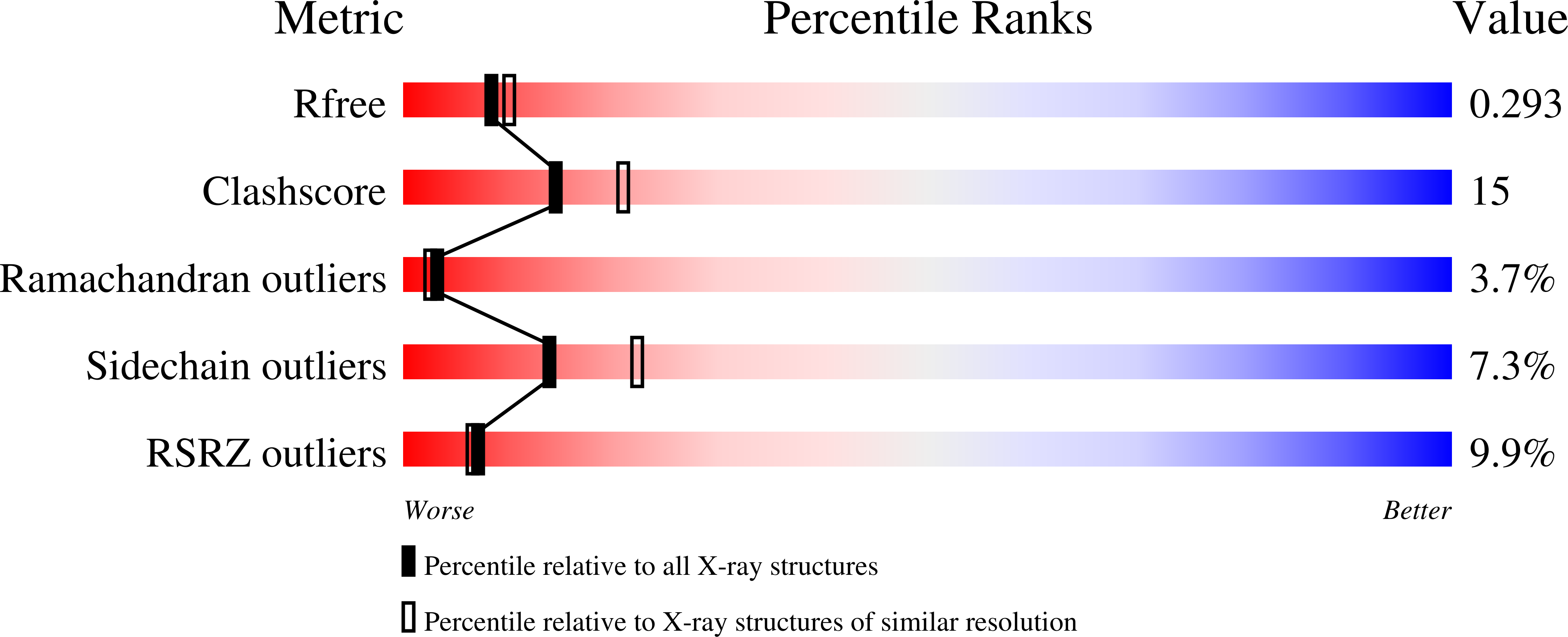
  • R-Value Free: 0.294 
  • R-Value Work: 0.242 
  • R-Value Observed: 0.246 

Starting Model: experimental
View more details

wwPDB Validation   3D Report Full Report


Ligand Structure Quality Assessment 


This is version 1.5 of the entry. See complete history


Literature

Structural and Mutational Analyses of a CD8alphabeta Heterodimer and Comparison with the CD8alphaalpha Homodimer.

Chang, H.C.Tan, K.Ouyang, J.Parisini, E.Liu, J.H.Le, Y.Wang, X.Reinherz, E.L.Wang, J.H.

(2005) Immunity 23: 661-671

  • DOI: https://doi.org/10.1016/j.immuni.2005.11.002
  • Primary Citation of Related Structures:  
    2ATP

  • PubMed Abstract: 

    The crystal structure of a recombinant mouse single chain CD8alphabeta ectodomains at 2.4 A resolution reveals paired immunoglobulin variable region-like domains with a striking resemblance to CD8alphaalpha in size, shape, and surface electrostatic potential of complementarity-determining regions (CDR), despite <20% sequence identity between the CD8alpha and CD8beta subunits. Unlike the CD8alpha subunit(s) in the heterodimer or homodimer, the CDR1 loop of CD8beta tilts away from its corresponding CDR2 and CDR3 loops. Consistent with this observation, independent mutational studies reveal that alanine substitutions of residues in the CDR1 loop of CD8beta have no effect on CD8alphabeta coreceptor function, whereas mutations in CD8beta CDR2 and CDR3 loops abolish CD8alphabeta coreceptor activity. The implications of these findings and additional CD8alpha mutational studies for CD8alphabeta- versus CD8alphaalpha-MHCI binding are discussed.


  • Organizational Affiliation

    Laboratory of Immunobiology, Department of Medical Oncology, Dana-Farber Cancer Institute, Department of Medicine, Harvard Medical School, Boston, Massachusetts 02115, USA. hsiu-ching_chang@dfci.harvard.edu


Macromolecules
Find similar proteins by:  (by identity cutoff)  |  3D Structure
Entity ID: 1
MoleculeChains Sequence LengthOrganismDetailsImage
T-cell surface glycoprotein CD8 alpha chainA,
D [auth C]
122Mus musculusMutation(s): 0 
Gene Names: Cd8aLyt-2Lyt2
UniProt
Find proteins for P01731 (Mus musculus)
Explore P01731 
Go to UniProtKB:  P01731
Entity Groups  
Sequence Clusters30% Identity50% Identity70% Identity90% Identity95% Identity100% Identity
UniProt GroupP01731
Glycosylation
Glycosylation Sites: 1Go to GlyGen: P01731-1
Sequence Annotations
Expand
  • Reference Sequence
Find similar proteins by:  (by identity cutoff)  |  3D Structure
Entity ID: 2
MoleculeChains Sequence LengthOrganismDetailsImage
artifact linkerB [auth E],
E [auth F]
29N/AMutation(s): 0 
Entity Groups  
Sequence Clusters30% Identity50% Identity70% Identity90% Identity95% Identity100% Identity
Sequence Annotations
Expand
  • Reference Sequence
Find similar proteins by:  (by identity cutoff)  |  3D Structure
Entity ID: 3
MoleculeChains Sequence LengthOrganismDetailsImage
T-cell surface glycoprotein CD8 beta chainC [auth B],
F [auth D]
115Mus musculusMutation(s): 0 
Gene Names: Cd8bLyt-3Lyt3
UniProt
Find proteins for P10300 (Mus musculus)
Explore P10300 
Go to UniProtKB:  P10300
Entity Groups  
Sequence Clusters30% Identity50% Identity70% Identity90% Identity95% Identity100% Identity
UniProt GroupP10300
Glycosylation
Glycosylation Sites: 1Go to GlyGen: P10300-1
Sequence Annotations
Expand
  • Reference Sequence
Experimental Data & Validation

Experimental Data

  • Method: X-RAY DIFFRACTION
  • Resolution: 2.40 Å
  • R-Value Free: 0.294 
  • R-Value Work: 0.242 
  • R-Value Observed: 0.246 
  • Space Group: P 1 21 1
Unit Cell:
Length ( Å )Angle ( ˚ )
a = 37.668α = 90
b = 92.655β = 95.67
c = 79.33γ = 90
Software Package:
Software NamePurpose
HKL-2000data collection
SCALEPACKdata scaling
PHASESphasing
CNSrefinement
HKL-2000data reduction

Structure Validation

View Full Validation Report


Ligand Structure Quality Assessment 


Entry History 

Deposition Data

Revision History  (Full details and data files)

  • Version 1.0: 2005-12-27
    Type: Initial release
  • Version 1.1: 2008-04-30
    Changes: Version format compliance
  • Version 1.2: 2011-07-13
    Changes: Non-polymer description, Version format compliance
  • Version 1.3: 2020-07-29
    Type: Remediation
    Reason: Carbohydrate remediation
    Changes: Data collection, Derived calculations, Structure summary
  • Version 1.4: 2023-08-23
    Changes: Data collection, Database references, Refinement description, Structure summary
  • Version 1.5: 2024-10-30
    Changes: Structure summary