2DU1

Crystal Structure of basic winged bean lectin in complex with Methyl-alpha-N-acetyl-D galactosamine


Experimental Data Snapshot

  • Method: X-RAY DIFFRACTION
  • Resolution: 2.60 Å
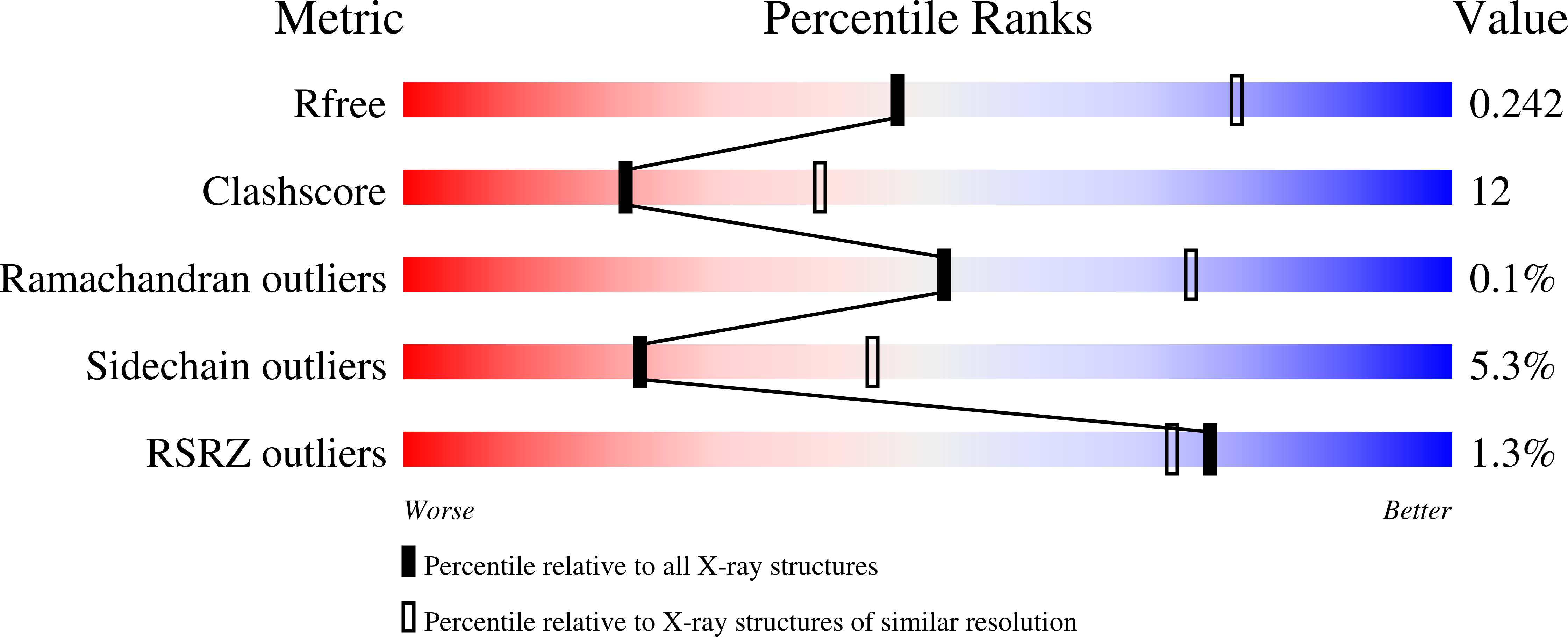
  • R-Value Free: 0.251 
  • R-Value Work: 0.194 
  • R-Value Observed: 0.194 

Starting Model: experimental
View more details

wwPDB Validation   3D Report Full Report


Ligand Structure Quality Assessment 


This is version 2.1 of the entry. See complete history


Literature

Structural basis for the carbohydrate-specificity of basic winged-bean lectin and its differential affinity for Gal and GalNAc

Kulkarni, K.A.Katiyar, S.Surolia, A.Vijayan, M.Suguna, K.

(2006) Acta Crystallogr D Biol Crystallogr 62: 1319-1324

  • DOI: https://doi.org/10.1107/S0907444906028198
  • Primary Citation of Related Structures:  
    2DTW, 2DTY, 2DU0, 2DU1

  • PubMed Abstract: 

    The crystal structure of the complexes of basic winged-bean lectin with galactose, 2-methoxygalactose, N-acetylgalactosamine and methyl-alpha-N-acetylgalactosamine have been determined. Lectin-sugar interactions involve four hydrogen bonds and a stacking interaction in all of the complexes. In addition, an N-H...O hydrogen bond involving the hydroxyl group at C2 exists in the galactose and 2-methoxygalactose complexes. An additional hydrophobic interaction involving the methyl group in the latter leads to the higher affinity of the methyl derivative. In the lectin-N-acetylgalactosamine complex the N-H...O hydrogen bond is lost, but a compensatory hydrogen bond is formed involving the O atom of the acetamido group. In addition, the CH(3) moiety of the acetamido group is involved in hydrophobic interactions. Consequently, the 2-methyl and acetamido derivatives of galactose have nearly the same affinity for the lectin. The methyl group alpha-linked to the galactose takes part in additional hydrophobic interactions. Therefore, methyl-alpha-N-acetylgalactosamine has a higher affinity than N-acetylgalactosamine for the lectin. The structures of basic winged-bean lectin-sugar complexes provide a framework for examining the relative affinity of galactose and galactosamine for the lectins that bind to them. The complexes also lead to a structural explanation for the blood-group specificity of basic winged-bean lectin.


  • Organizational Affiliation

    Molecular Biophysics Unit, Indian Institute of Science, Bangalore, India.


Macromolecules
Find similar proteins by:  (by identity cutoff)  |  3D Structure
Entity ID: 1
MoleculeChains Sequence LengthOrganismDetailsImage
Basic agglutinin
A, B, C, D
241Psophocarpus tetragonolobusMutation(s): 0 
UniProt
Find proteins for O24313 (Psophocarpus tetragonolobus)
Explore O24313 
Go to UniProtKB:  O24313
Entity Groups  
Sequence Clusters30% Identity50% Identity70% Identity90% Identity95% Identity100% Identity
UniProt GroupO24313
Glycosylation
Glycosylation Sites: 2
Sequence Annotations
Expand
  • Reference Sequence
Oligosaccharides

Help

Entity ID: 2
MoleculeChains Length2D Diagram Glycosylation3D Interactions
alpha-L-fucopyranose-(1-3)-[2-acetamido-2-deoxy-beta-D-glucopyranose-(1-4)]2-acetamido-2-deoxy-beta-D-glucopyranose
E, F, G, H
3N-Glycosylation
Glycosylation Resources
GlyTouCan:  G11971MR
GlyCosmos:  G11971MR
GlyGen:  G11971MR
Entity ID: 3
MoleculeChains Length2D Diagram Glycosylation3D Interactions
2-acetamido-2-deoxy-beta-D-glucopyranose-(1-4)-2-acetamido-2-deoxy-beta-D-glucopyranose
I
2N-Glycosylation
Glycosylation Resources
GlyTouCan:  G42666HT
GlyCosmos:  G42666HT
GlyGen:  G42666HT
Small Molecules
Ligands 4 Unique
IDChains Name / Formula / InChI Key2D Diagram3D Interactions
MGC
Query on MGC

Download Ideal Coordinates CCD File 
J [auth A],
N [auth B],
Q [auth C],
U [auth D]
methyl 2-acetamido-2-deoxy-alpha-D-galactopyranoside
C9 H17 N O6
ZEVOCXOZYFLVKN-ZEBDFXRSSA-N
NAG
Query on NAG

Download Ideal Coordinates CCD File 
K [auth A],
R [auth C],
V [auth D]
2-acetamido-2-deoxy-beta-D-glucopyranose
C8 H15 N O6
OVRNDRQMDRJTHS-FMDGEEDCSA-N
MN
Query on MN

Download Ideal Coordinates CCD File 
M [auth A],
P [auth B],
T [auth C],
X [auth D]
MANGANESE (II) ION
Mn
WAEMQWOKJMHJLA-UHFFFAOYSA-N
CA
Query on CA

Download Ideal Coordinates CCD File 
L [auth A],
O [auth B],
S [auth C],
W [auth D]
CALCIUM ION
Ca
BHPQYMZQTOCNFJ-UHFFFAOYSA-N
Experimental Data & Validation

Experimental Data

  • Method: X-RAY DIFFRACTION
  • Resolution: 2.60 Å
  • R-Value Free: 0.251 
  • R-Value Work: 0.194 
  • R-Value Observed: 0.194 
  • Space Group: P 21 21 2
Unit Cell:
Length ( Å )Angle ( ˚ )
a = 157.619α = 90
b = 90.96β = 90
c = 73.43γ = 90
Software Package:
Software NamePurpose
CNSrefinement
HKL-2000data reduction
SCALEPACKdata scaling
CNSphasing

Structure Validation

View Full Validation Report


Ligand Structure Quality Assessment 


Entry History 

Deposition Data

Revision History  (Full details and data files)

  • Version 1.0: 2006-11-07
    Type: Initial release
  • Version 1.1: 2008-04-30
    Changes: Version format compliance
  • Version 1.2: 2011-07-13
    Changes: Non-polymer description, Version format compliance
  • Version 1.3: 2017-10-11
    Changes: Data collection
  • Version 2.0: 2020-07-29
    Type: Remediation
    Reason: Carbohydrate remediation
    Changes: Atomic model, Data collection, Derived calculations, Structure summary
  • Version 2.1: 2023-10-25
    Changes: Data collection, Database references, Refinement description, Structure summary