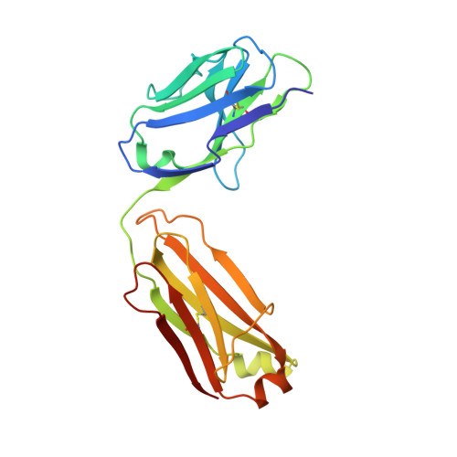
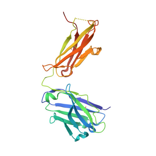
2FJF

Structure of the G6 Fab, a phage derived VEGF binding Fab


Experimental Data Snapshot

  • Method: X-RAY DIFFRACTION
  • Resolution: 2.65 Å
  • R-Value Free: 0.248 
  • R-Value Work: 0.199 
  • R-Value Observed: 0.200 

wwPDB Validation   3D Report Full Report


This is version 1.3 of the entry. See complete history


Literature

Structure-function studies of two synthetic anti-vascular endothelial growth factor Fabs and comparison with the Avastin Fab.

Fuh, G.Wu, P.Liang, W.C.Ultsch, M.Lee, C.V.Moffat, B.Wiesmann, C.

(2006) J Biol Chem 281: 6625-6631

  • DOI: https://doi.org/10.1074/jbc.M507783200
  • Primary Citation of Related Structures:  
    2FJF, 2FJG, 2FJH

  • PubMed Abstract: 

    In the quest to discover new research tools and to develop better agents in the fight against cancer, two antibodies, G6 and B20-4, were isolated from synthetic antibody phage libraries. Unlike the AVASTINtrade mark antibody, a recently approved agent for the treatment of patients with colorectal cancer, B20-4 and G6 bind and block both human and murine vascular endothelial growth factor (VEGF). Here we have analyzed and compared the binding epitopes on VEGF for these three antibodies using alanine-scanning mutagenesis and structural analyses. The epitopes recognized by both synthetic antibodies are conserved between human and mouse VEGF, and they match closely to the receptor epitopes both structurally and functionally. In contrast, the Avastin epitope overlaps minimally with the receptor binding surface and centers around a residue that is not conserved in mouse. Our structural and functional analyses elucidate the cross-species reactivity of all three antibodies and emphasize the potential advantages of antibody generation using phage display as the resulting antibodies do not depend on sequence differences across species and preferentially target natural protein-protein interaction surfaces.


  • Organizational Affiliation

    Department of Protein Engineering, Genentech, Inc., South San Francisco, California 94080, USA. gml@gene.com


Macromolecules
Find similar proteins by:  (by identity cutoff)  |  3D Structure
Entity ID: 1
MoleculeChains Sequence LengthOrganismDetailsImage
Light Chain of a VEGF binding Antibody214Homo sapiensMutation(s): 0 
Entity Groups  
Sequence Clusters30% Identity50% Identity70% Identity90% Identity95% Identity100% Identity
Sequence Annotations
Expand
  • Reference Sequence
Find similar proteins by:  (by identity cutoff)  |  3D Structure
Entity ID: 2
MoleculeChains Sequence LengthOrganismDetailsImage
Heavy Chain of a VEGF binding Antibody227Homo sapiensMutation(s): 0 
Entity Groups  
Sequence Clusters30% Identity50% Identity70% Identity90% Identity95% Identity100% Identity
Sequence Annotations
Expand
  • Reference Sequence
Experimental Data & Validation

Experimental Data

  • Method: X-RAY DIFFRACTION
  • Resolution: 2.65 Å
  • R-Value Free: 0.248 
  • R-Value Work: 0.199 
  • R-Value Observed: 0.200 
  • Space Group: C 1 2 1
Unit Cell:
Length ( Å )Angle ( ˚ )
a = 274.919α = 90
b = 192.229β = 117
c = 154.108γ = 90
Software Package:
Software NamePurpose
REFMACrefinement
HKL-2000data reduction
SCALEPACKdata scaling
AMoREphasing

Structure Validation

View Full Validation Report


Entry History 

Deposition Data

  • Released Date: 2006-02-07 
  • Deposition Author(s): Wiesmann, C.

Revision History  (Full details and data files)

  • Version 1.0: 2006-02-07
    Type: Initial release
  • Version 1.1: 2008-05-01
    Changes: Version format compliance
  • Version 1.2: 2011-07-13
    Changes: Advisory, Version format compliance
  • Version 1.3: 2024-10-09
    Changes: Data collection, Database references, Refinement description, Structure summary