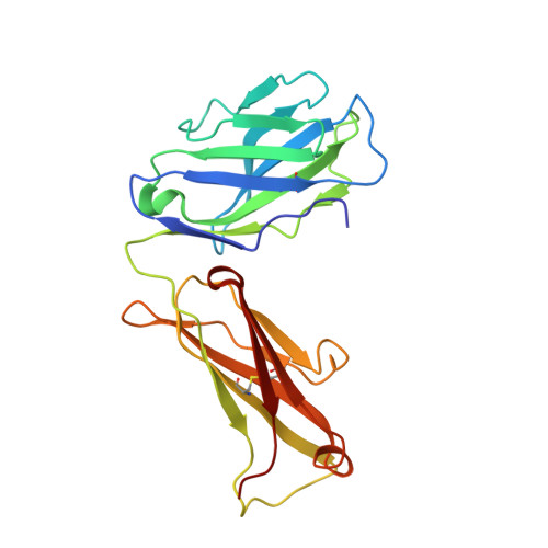
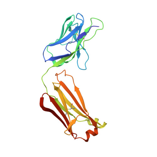
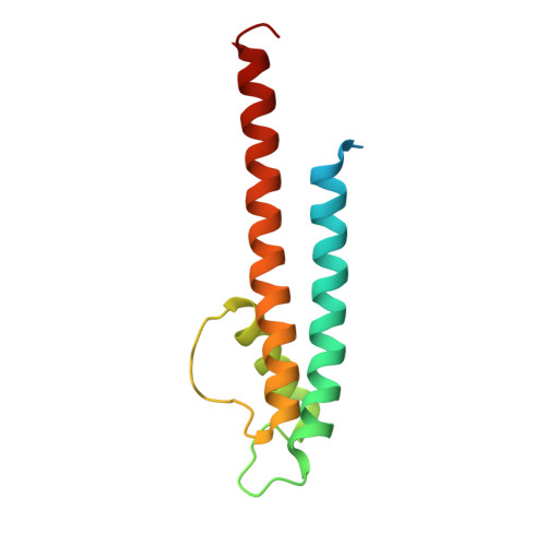
2HVJ

Crystal structure of KcsA-Fab-TBA complex in low K+


Experimental Data Snapshot

  • Method: X-RAY DIFFRACTION
  • Resolution: 2.75 Å
  • R-Value Free: 0.246 
  • R-Value Work: 0.222 
  • R-Value Observed: 0.222 

Starting Model: experimental
View more details

wwPDB Validation   3D Report Full Report


Ligand Structure Quality Assessment 


This is version 1.7 of the entry. See complete history


Literature

Crystallographic Study of the Tetrabutylammonium Block to the KcsA K(+) Channel.

Yohannan, S.Hu, Y.Zhou, Y.

(2007) J Mol Biol 366: 806-814

  • DOI: https://doi.org/10.1016/j.jmb.2006.11.081
  • Primary Citation of Related Structures:  
    2DWD, 2DWE, 2HVJ, 2HVK

  • PubMed Abstract: 

    K(+) channels play essential roles in regulating membrane excitability of many diverse cell types by selectively conducting K(+) ions through their pores. Many diverse molecules can plug the pore and modulate the K(+) current. Quaternary ammonium (QA) ions are a class of pore blockers that have been used for decades by biophysicists to probe the pore, leading to important insights into the structure-function relation of K(+) channels. However, many key aspects of the QA-blocking mechanisms remain unclear to date, and understanding these questions requires high resolution structural information. Here, we address the question of whether intracellular QA blockade causes conformational changes of the K(+) channel selectivity filter. We have solved the structures of the KcsA K(+) channel in complex with tetrabutylammonium (TBA) and tetrabutylantimony (TBSb) under various ionic conditions. Our results demonstrate that binding of TBA or TBSb causes no significant change in the KcsA structure at high concentrations of permeant ions. We did observe the expected conformational change of the filter at low concentration of K(+), but this change appears to be independent of TBA or TBSb blockade.


  • Organizational Affiliation

    Department of Cellular and Molecular Physiology, Yale University School of Medicine, New Haven, CT 06520, USA.


Macromolecules
Find similar proteins by:  (by identity cutoff)  |  3D Structure
Entity ID: 1
MoleculeChains Sequence LengthOrganismDetailsImage
antibody Fab heavy chain219Mus musculusMutation(s): 0 
Entity Groups  
Sequence Clusters30% Identity50% Identity70% Identity90% Identity95% Identity100% Identity
Sequence Annotations
Expand
  • Reference Sequence
Find similar proteins by:  (by identity cutoff)  |  3D Structure
Entity ID: 2
MoleculeChains Sequence LengthOrganismDetailsImage
antibody Fab light chain212Mus musculusMutation(s): 0 
Entity Groups  
Sequence Clusters30% Identity50% Identity70% Identity90% Identity95% Identity100% Identity
Sequence Annotations
Expand
  • Reference Sequence
Find similar proteins by:  (by identity cutoff)  |  3D Structure
Entity ID: 3
MoleculeChains Sequence LengthOrganismDetailsImage
Voltage-gated potassium channel124Streptomyces lividansMutation(s): 2 
Gene Names: kcsA
Membrane Entity: Yes 
UniProt
Find proteins for P0A334 (Streptomyces lividans)
Explore P0A334 
Go to UniProtKB:  P0A334
Entity Groups  
Sequence Clusters30% Identity50% Identity70% Identity90% Identity95% Identity100% Identity
UniProt GroupP0A334
Sequence Annotations
Expand
  • Reference Sequence
Small Molecules
Ligands 4 Unique
IDChains Name / Formula / InChI Key2D Diagram3D Interactions
L2C
Query on L2C

Download Ideal Coordinates CCD File 
G [auth C](2S)-3-HYDROXY-2-(NONANOYLOXY)PROPYL LAURATE
C24 H46 O5
MYFVXYUACRBWGD-QFIPXVFZSA-N
TBA
Query on TBA

Download Ideal Coordinates CCD File 
H [auth C]TETRABUTYLAMMONIUM ION
C16 H36 N
DZLFLBLQUQXARW-UHFFFAOYSA-N
F09
Query on F09

Download Ideal Coordinates CCD File 
D [auth A]NONAN-1-OL
C9 H20 O
ZWRUINPWMLAQRD-UHFFFAOYSA-N
K
Query on K

Download Ideal Coordinates CCD File 
E [auth C],
F [auth C]
POTASSIUM ION
K
NPYPAHLBTDXSSS-UHFFFAOYSA-N
Experimental Data & Validation

Experimental Data

  • Method: X-RAY DIFFRACTION
  • Resolution: 2.75 Å
  • R-Value Free: 0.246 
  • R-Value Work: 0.222 
  • R-Value Observed: 0.222 
  • Space Group: I 4
Unit Cell:
Length ( Å )Angle ( ˚ )
a = 156.423α = 90
b = 156.423β = 90
c = 75.797γ = 90
Software Package:
Software NamePurpose
CNSrefinement
SCALEPACKdata scaling
REFMACrefinement
PDB_EXTRACTdata extraction
HKL-2000data reduction
CNSphasing
DENZOdata reduction

Structure Validation

View Full Validation Report


Ligand Structure Quality Assessment 


Entry History 

Deposition Data

  • Released Date: 2007-02-20 
  • Deposition Author(s): Zhou, Y.

Revision History  (Full details and data files)

  • Version 1.0: 2007-02-20
    Type: Initial release
  • Version 1.1: 2008-05-01
    Changes: Version format compliance
  • Version 1.2: 2011-07-13
    Changes: Derived calculations, Version format compliance
  • Version 1.3: 2017-10-18
    Changes: Refinement description
  • Version 1.4: 2019-07-24
    Changes: Data collection, Refinement description
  • Version 1.5: 2021-10-20
    Changes: Database references, Derived calculations
  • Version 1.6: 2023-08-30
    Changes: Data collection, Refinement description
  • Version 1.7: 2024-11-20
    Changes: Structure summary