2OYE

Indomethacin-(R)-alpha-ethyl-ethanolamide bound to Cyclooxygenase-1


Experimental Data Snapshot

  • Method: X-RAY DIFFRACTION
  • Resolution: 2.85 Å
  • R-Value Free: 
    0.292 (Depositor), 0.283 (DCC) 
  • R-Value Work: 
    0.245 (Depositor), 0.239 (DCC) 

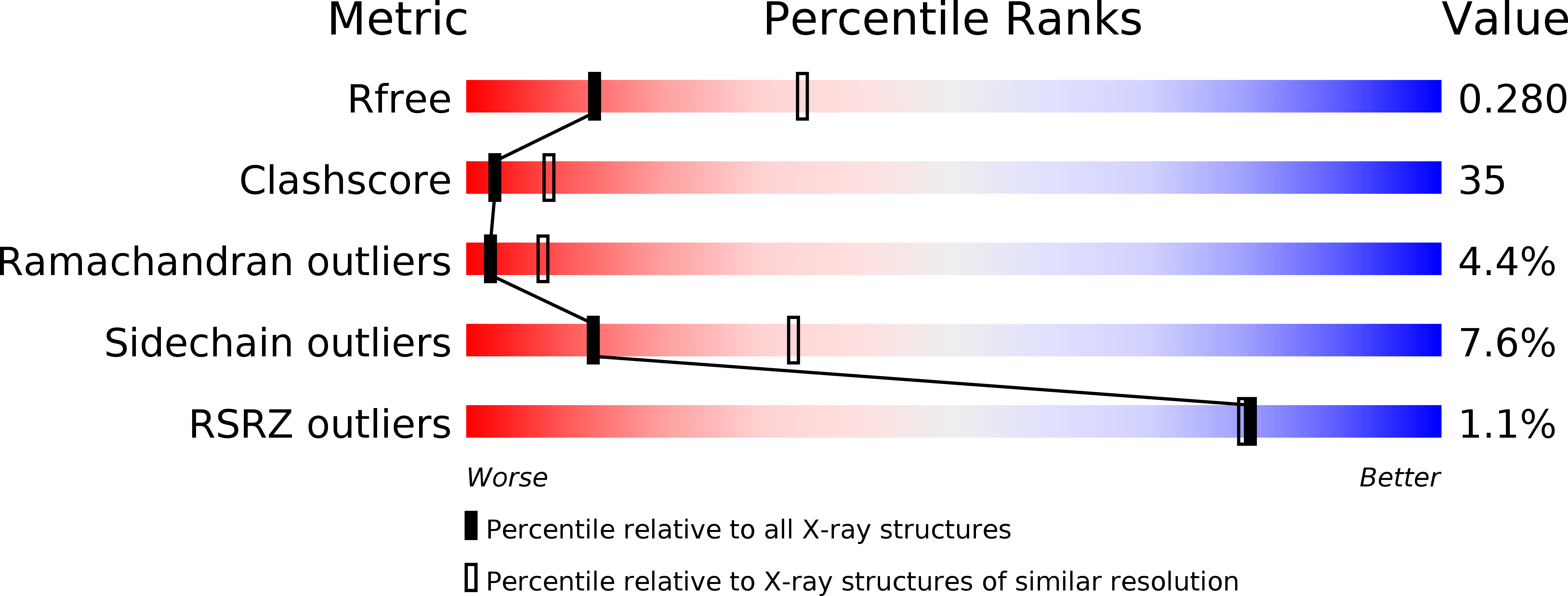
wwPDB Validation   3D Report Full Report


Ligand Structure Quality Assessment 


This is version 2.1 of the entry. See complete history


Literature

Structural basis of enantioselective inhibition of cyclooxygenase-1 by S-alpha-substituted indomethacin ethanolamides.

Harman, C.A.Turman, M.V.Kozak, K.R.Marnett, L.J.Smith, W.L.Garavito, R.M.

(2007) J Biol Chem 282: 28096-28105

  • DOI: https://doi.org/10.1074/jbc.M701335200
  • Primary Citation of Related Structures:  
    2OYE, 2OYU

  • PubMed Abstract: 

    The modification of the nonselective nonsteroidal anti-inflammatory drug, indomethacin, by amidation presents a promising strategy for designing novel cyclooxygenase (COX)-2-selective inhibitors. A series of alpha-substituted indomethacin ethanolamides, which exist as R/S-enantiomeric pairs, provides a means to study the impact of stereochemistry on COX inhibition. Comparative studies revealed that the R- and S-enantiomers of the alpha-substituted analogs inhibit COX-2 with almost equal efficacy, whereas COX-1 is selectively inhibited by the S-enantiomers. Mutagenesis studies have not been able to identify residues that manifest the enantioselectivity in COX-1. In an effort to understand the structural impact of chirality on COX-1 selectivity, the crystal structures of ovine COX-1 in complexes with an enantiomeric pair of these indomethacin ethanolamides were determined at resolutions between 2.75 and 2.85 A. These structures reveal unique, enantiomer-selective interactions within the COX-1 side pocket region that stabilize drug binding and account for the chiral selectivity observed with the (S)-alpha-substituted indomethacin ethanolamides. Kinetic analysis of binding demonstrates that both inhibitors bind quickly utilizing a two-step mechanism. However, the second binding step is readily reversible for the R-enantiomer, whereas for the S-enantiomer, it is not. These studies establish for the first time the structural and kinetic basis of high affinity binding of a neutral inhibitor to COX-1 and demonstrate that the side pocket of COX-1, previously thought to be sterically inaccessible, can serve as a binding pocket for inhibitor association.


  • Organizational Affiliation

    Department of Biochemistry and Molecular Biology, Michigan State University, East Lansing, Michigan 48824, USA.


Macromolecules
Find similar proteins by:  (by identity cutoff)  |  3D Structure
Entity ID: 1
MoleculeChains Sequence LengthOrganismDetailsImage
Prostaglandin G/H synthase 1A [auth P]600Ovis ariesMutation(s): 0 
EC: 1.14.99.1
UniProt
Find proteins for P05979 (Ovis aries)
Explore P05979 
Go to UniProtKB:  P05979
Entity Groups  
Sequence Clusters30% Identity50% Identity70% Identity90% Identity95% Identity100% Identity
UniProt GroupP05979
Glycosylation
Glycosylation Sites: 3
Sequence Annotations
Expand
  • Reference Sequence
Oligosaccharides

Help

Entity ID: 2
MoleculeChains Length2D Diagram Glycosylation3D Interactions
2-acetamido-2-deoxy-alpha-D-glucopyranose-(1-4)-2-acetamido-2-deoxy-beta-D-glucopyranoseB [auth A]2N-Glycosylation
Glycosylation Resources
GlyTouCan:  G07375KG
GlyCosmos:  G07375KG
GlyGen:  G07375KG
Entity ID: 3
MoleculeChains Length2D Diagram Glycosylation3D Interactions
beta-D-mannopyranose-(1-6)-beta-D-mannopyranose-(1-4)-2-acetamido-2-deoxy-beta-D-glucopyranose-(1-4)-2-acetamido-2-deoxy-beta-D-glucopyranoseC [auth B]4N-Glycosylation
Glycosylation Resources
GlyTouCan:  G40894EI
GlyCosmos:  G40894EI
GlyGen:  G40894EI
Entity ID: 4
MoleculeChains Length2D Diagram Glycosylation3D Interactions
2-acetamido-2-deoxy-beta-D-glucopyranose-(1-4)-2-acetamido-2-deoxy-beta-D-glucopyranoseD [auth C]2N-Glycosylation
Glycosylation Resources
GlyTouCan:  G42666HT
GlyCosmos:  G42666HT
GlyGen:  G42666HT
Small Molecules
Ligands 4 Unique
IDChains Name / Formula / InChI Key2D Diagram3D Interactions
HEM
Query on HEM

Download Ideal Coordinates CCD File 
H [auth P]PROTOPORPHYRIN IX CONTAINING FE
C34 H32 Fe N4 O4
KABFMIBPWCXCRK-RGGAHWMASA-L
IM8
Query on IM8

Download Ideal Coordinates CCD File 
I [auth P]2-[1-(4-CHLOROBENZOYL)-5-METHOXY-2-METHYL-1H-INDOL-3-YL]-N-[(1R)-1-(HYDROXYMETHYL)PROPYL]ACETAMIDE
C23 H25 Cl N2 O4
GKJWXEORYGBJFS-QGZVFWFLSA-N
BOG
Query on BOG

Download Ideal Coordinates CCD File 
E [auth P],
F [auth P]
octyl beta-D-glucopyranoside
C14 H28 O6
HEGSGKPQLMEBJL-RKQHYHRCSA-N
FLC
Query on FLC

Download Ideal Coordinates CCD File 
G [auth P]CITRATE ANION
C6 H5 O7
KRKNYBCHXYNGOX-UHFFFAOYSA-K
Binding Affinity Annotations 
IDSourceBinding Affinity
IM8 BindingDB:  2OYE IC50: 5.90e+4 (nM) from 1 assay(s)
Experimental Data & Validation

Experimental Data

  • Method: X-RAY DIFFRACTION
  • Resolution: 2.85 Å
  • R-Value Free:  0.292 (Depositor), 0.283 (DCC) 
  • R-Value Work:  0.245 (Depositor), 0.239 (DCC) 
Space Group: P 65 2 2
Unit Cell:
Length ( Å )Angle ( ˚ )
a = 181.72α = 90
b = 181.72β = 90
c = 104.09γ = 120
Software Package:
Software NamePurpose
DENZOdata reduction
SCALEPACKdata scaling
CNSrefinement
PDB_EXTRACTdata extraction
MAR345data collection
CNSphasing

Structure Validation

View Full Validation Report


Ligand Structure Quality Assessment 


Entry History 

Deposition Data

Revision History  (Full details and data files)

  • Version 1.0: 2007-07-24
    Type: Initial release
  • Version 1.1: 2007-10-03
    Changes: Version format compliance
  • Version 1.2: 2011-07-13
    Changes: Derived calculations, Version format compliance
  • Version 1.3: 2017-10-18
    Changes: Advisory, Refinement description
  • Version 2.0: 2020-07-29
    Type: Remediation
    Reason: Carbohydrate remediation
    Changes: Advisory, Atomic model, Data collection, Derived calculations, Structure summary
  • Version 2.1: 2024-10-30
    Changes: Data collection, Database references, Structure summary