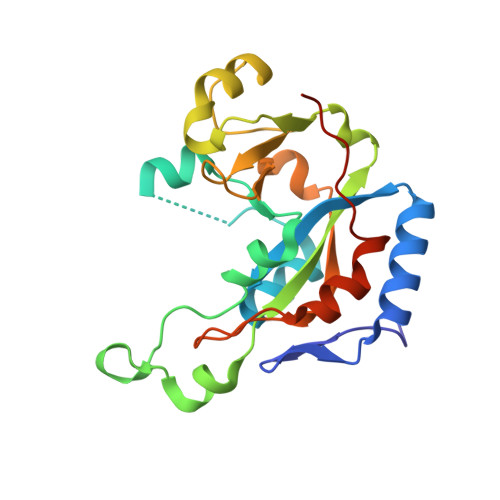
2Q6M

Catalytic fragment of Cholix toxin from Vibrio Cholerae in complex with the PJ34 inhibitor


Experimental Data Snapshot

  • Method: X-RAY DIFFRACTION
  • Resolution: 1.25 Å
  • R-Value Free: 0.180 
  • R-Value Work: 0.150 
  • R-Value Observed: 0.151 

wwPDB Validation   3D Report Full Report


Ligand Structure Quality Assessment 


This is version 1.3 of the entry. See complete history


Literature

Cholix Toxin, a Novel ADP-ribosylating Factor from Vibrio cholerae.

Jorgensen, R.Purdy, A.E.Fieldhouse, R.J.Kimber, M.S.Bartlett, D.H.Merrill, A.R.

(2008) J Biol Chem 283: 10671-10678

  • DOI: https://doi.org/10.1074/jbc.M710008200
  • Primary Citation of Related Structures:  
    2Q5T, 2Q6M

  • PubMed Abstract: 

    The ADP-ribosyltransferases are a class of enzymes that display activity in a variety of bacterial pathogens responsible for causing diseases in plants and animals, including those affecting mankind, such as diphtheria, cholera, and whooping cough. We report the characterization of a novel toxin from Vibrio cholerae, which we call cholix toxin. The toxin is active against mammalian cells (IC(50) = 4.6 +/- 0.4 ng/ml) and crustaceans (Artemia nauplii LD(50) = 10 +/- 2 mug/ml). Here we show that this toxin is the third member of the diphthamide-specific class of ADP-ribose transferases and that it possesses specific ADP-ribose transferase activity against ribosomal eukaryotic elongation factor 2. We also describe the high resolution crystal structures of the multidomain toxin and its catalytic domain at 2.1- and 1.25-A resolution, respectively. The new structural data show that cholix toxin possesses the necessary molecular features required for infection of eukaryotes by receptor-mediated endocytosis, translocation to the host cytoplasm, and inhibition of protein synthesis by specific modification of elongation factor 2. The crystal structures also provide important insight into the structural basis for activation of toxin ADP-ribosyltransferase activity. These results indicate that cholix toxin may be an important virulence factor of Vibrio cholerae that likely plays a significant role in the survival of the organism in an aquatic environment.


  • Organizational Affiliation

    Department of Molecular and Cellular Biology, University of Guelph, Guelph, Ontario N1G 2W1, Canada.


Macromolecules
Find similar proteins by:  (by identity cutoff)  |  3D Structure
Entity ID: 1
MoleculeChains Sequence LengthOrganismDetailsImage
Cholix toxin212Vibrio choleraeMutation(s): 0 
Gene Names: toxA
EC: 2.4.2.36
UniProt
Find proteins for Q5EK40 (Vibrio cholerae)
Explore Q5EK40 
Go to UniProtKB:  Q5EK40
Entity Groups  
Sequence Clusters30% Identity50% Identity70% Identity90% Identity95% Identity100% Identity
UniProt GroupQ5EK40
Sequence Annotations
Expand
  • Reference Sequence
Small Molecules
Ligands 1 Unique
IDChains Name / Formula / InChI Key2D Diagram3D Interactions
P34
Query on P34

Download Ideal Coordinates CCD File 
B [auth A],
C [auth A]
N~2~,N~2~-DIMETHYL-N~1~-(6-OXO-5,6-DIHYDROPHENANTHRIDIN-2-YL)GLYCINAMIDE
C17 H17 N3 O2
UYJZZVDLGDDTCL-UHFFFAOYSA-N
Experimental Data & Validation

Experimental Data

  • Method: X-RAY DIFFRACTION
  • Resolution: 1.25 Å
  • R-Value Free: 0.180 
  • R-Value Work: 0.150 
  • R-Value Observed: 0.151 
  • Space Group: P 21 21 21
Unit Cell:
Length ( Å )Angle ( ˚ )
a = 40.085α = 90
b = 64.756β = 90
c = 77.995γ = 90
Software Package:
Software NamePurpose
DENZOdata reduction
SCALEPACKdata scaling
SOLVEphasing
RESOLVEphasing
REFMACrefinement
PDB_EXTRACTdata extraction
HKL-3000data collection
HKL-3000data reduction

Structure Validation

View Full Validation Report


Ligand Structure Quality Assessment 


Entry History 

Deposition Data

Revision History  (Full details and data files)

  • Version 1.0: 2008-02-12
    Type: Initial release
  • Version 1.1: 2011-07-13
    Changes: Version format compliance
  • Version 1.2: 2017-10-18
    Changes: Refinement description
  • Version 1.3: 2024-02-21
    Changes: Data collection, Database references, Derived calculations