2RF7

Crystal structure of the escherichia coli nrfa mutant Q263E


Experimental Data Snapshot

  • Method: X-RAY DIFFRACTION
  • Resolution: 2.04 Å
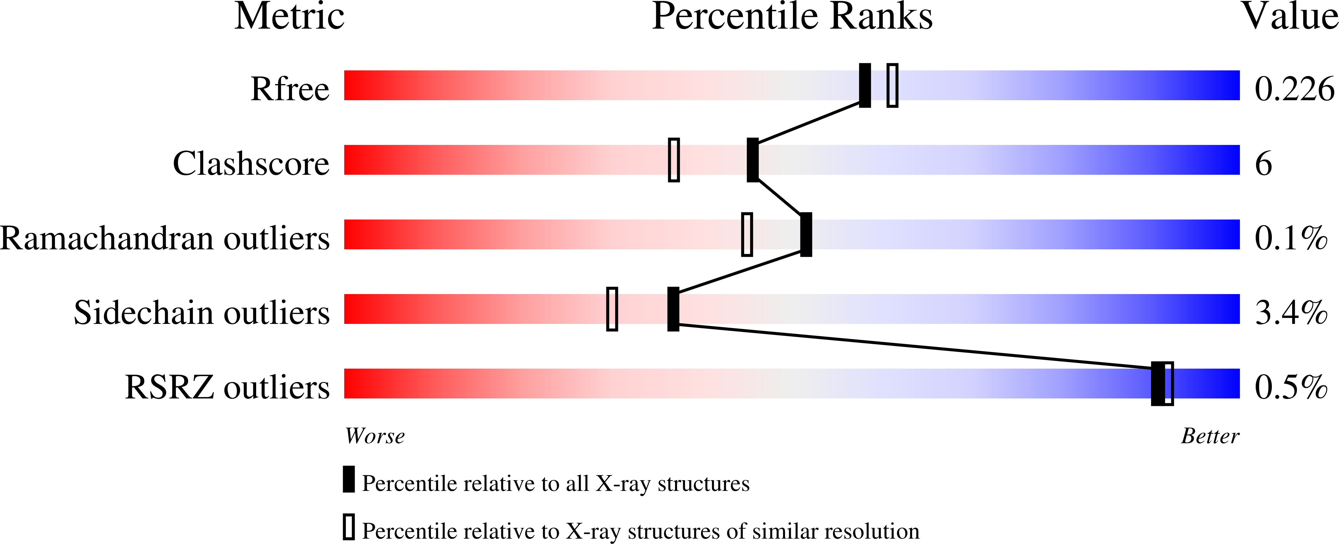
  • R-Value Free: 0.226 
  • R-Value Work: 0.171 
  • R-Value Observed: 0.174 

Starting Model: experimental
View more details

wwPDB Validation   3D Report Full Report


Ligand Structure Quality Assessment 


This is version 1.3 of the entry. See complete history


Literature

Role of a Conserved Glutamine Residue in Tuning the Catalytic Activity of Escherichia coli Cytochrome c Nitrite Reductase.

Clarke, T.A.Kemp, G.L.Wonderen, J.H.Doyle, R.M.Cole, J.A.Tovell, N.Cheesman, M.R.Butt, J.N.Richardson, D.J.Hemmings, A.M.

(2008) Biochemistry 47: 3789-3799

  • DOI: https://doi.org/10.1021/bi702175w
  • Primary Citation of Related Structures:  
    2RDZ, 2RF7

  • PubMed Abstract: 

    The pentaheme cytochrome c nitrite reductase (NrfA) of Escherichia coli is responsible for nitrite reduction during anaerobic respiration when nitrate is scarce. The NrfA active site consists of a hexacoordinate high-spin heme with a lysine ligand on the proximal side and water/hydroxide or substrate on the distal side. There are four further highly conserved active site residues including a glutamine (Q263) positioned 8 A from the heme iron for which the side chain, unusually, coordinates a conserved, essential calcium ion. Mutation of this glutamine to the more usual calcium ligand, glutamate, results in an increase in the K m for nitrite by around 10-fold, while V max is unaltered. Protein film voltammetry showed that lower potentials were required to detect activity from NrfA Q263E when compared with native enzyme, consistent with the introduction of a negative charge into the vicinity of the active site heme. EPR and MCD spectroscopic studies revealed the high spin state of the active site to be preserved, indicating that a water/hydroxide molecule is still coordinated to the heme in the resting state of the enzyme. Comparison of the X-ray crystal structures of the as-prepared, oxidized native and mutant enzymes showed an increased bond distance between the active site heme Fe(III) iron and the distal ligand in the latter as well as changes to the structure and mobility of the active site water molecule network. These results suggest that an important function of the unusual Q263-calcium ion pair is to increase substrate affinity through its role in supporting a network of hydrogen bonded water molecules stabilizing the active site heme distal ligand.


  • Organizational Affiliation

    Centre for Molecular and Structural Biochemistry, School of Biological Sciences, University of East Anglia, Norwich NR4 7TJ, United Kingdom.


Macromolecules
Find similar proteins by:  (by identity cutoff)  |  3D Structure
Entity ID: 1
MoleculeChains Sequence LengthOrganismDetailsImage
Cytochrome c-552
A, B, C, D
441Escherichia coli K-12Mutation(s): 1 
Gene Names: nrfA
EC: 1.7.2.2
UniProt
Find proteins for P0ABK9 (Escherichia coli (strain K12))
Explore P0ABK9 
Go to UniProtKB:  P0ABK9
Entity Groups  
Sequence Clusters30% Identity50% Identity70% Identity90% Identity95% Identity100% Identity
UniProt GroupP0ABK9
Sequence Annotations
Expand
  • Reference Sequence
Small Molecules
Ligands 3 Unique
IDChains Name / Formula / InChI Key2D Diagram3D Interactions
HEC
Query on HEC

Download Ideal Coordinates CCD File 
EA [auth C]
FA [auth C]
G [auth A]
GA [auth C]
H [auth A]
EA [auth C],
FA [auth C],
G [auth A],
GA [auth C],
H [auth A],
HA [auth C],
I [auth A],
IA [auth C],
J [auth A],
K [auth A],
OA [auth D],
PA [auth D],
Q [auth B],
QA [auth D],
R [auth B],
RA [auth D],
S [auth B],
SA [auth D],
T [auth B],
U [auth B]
HEME C
C34 H34 Fe N4 O4
HXQIYSLZKNYNMH-LJNAALQVSA-N
EDO
Query on EDO

Download Ideal Coordinates CCD File 
AA [auth B]
BA [auth B]
JA [auth C]
KA [auth C]
L [auth A]
AA [auth B],
BA [auth B],
JA [auth C],
KA [auth C],
L [auth A],
LA [auth C],
M [auth A],
N [auth A],
TA [auth D],
UA [auth D],
V [auth B],
W [auth B],
X [auth B],
Y [auth B],
Z [auth B]
1,2-ETHANEDIOL
C2 H6 O2
LYCAIKOWRPUZTN-UHFFFAOYSA-N
CA
Query on CA

Download Ideal Coordinates CCD File 
CA [auth C]
DA [auth C]
E [auth A]
F [auth A]
MA [auth D]
CA [auth C],
DA [auth C],
E [auth A],
F [auth A],
MA [auth D],
NA [auth D],
O [auth B],
P [auth B]
CALCIUM ION
Ca
BHPQYMZQTOCNFJ-UHFFFAOYSA-N
Experimental Data & Validation

Experimental Data

  • Method: X-RAY DIFFRACTION
  • Resolution: 2.04 Å
  • R-Value Free: 0.226 
  • R-Value Work: 0.171 
  • R-Value Observed: 0.174 
  • Space Group: P 21 21 21
Unit Cell:
Length ( Å )Angle ( ˚ )
a = 82.292α = 90
b = 91.201β = 90
c = 295.486γ = 90
Software Package:
Software NamePurpose
MOSFLMdata reduction
SCALAdata scaling
MOLREPphasing
REFMACrefinement
PDB_EXTRACTdata extraction
ADSCdata collection

Structure Validation

View Full Validation Report


Ligand Structure Quality Assessment 


Entry History 

Deposition Data

Revision History  (Full details and data files)

  • Version 1.0: 2008-03-25
    Type: Initial release
  • Version 1.1: 2011-07-13
    Changes: Version format compliance
  • Version 1.2: 2021-10-20
    Changes: Database references, Derived calculations
  • Version 1.3: 2023-08-30
    Changes: Data collection, Refinement description