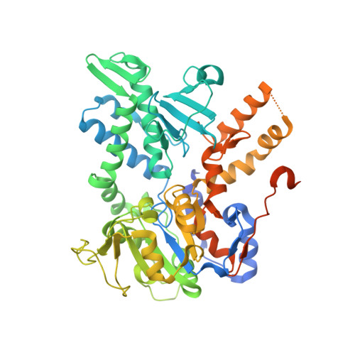
2XJD

Crystal structure of the D52N variant of cytosolic 5'-nucleotidase II in complex with inorganic phosphate and deoxyadenosine triphosphate


Experimental Data Snapshot

  • Method: X-RAY DIFFRACTION
  • Resolution: 2.00 Å
  • R-Value Free: 0.208 
  • R-Value Work: 0.176 
  • R-Value Observed: 0.178 

Starting Model: experimental
View more details

wwPDB Validation   3D Report Full Report


Ligand Structure Quality Assessment 


This is version 1.2 of the entry. See complete history


Literature

Structural Basis for the Allosteric Regulation and Substrate Recognition of Human Cytosolic 5'-Nucleotidase II.

Wallden, K.Nordlund, P.

(2011) J Mol Biol 408: 684

  • DOI: https://doi.org/10.1016/j.jmb.2011.02.059
  • Primary Citation of Related Structures:  
    2XCV, 2XCW, 2XCX, 2XJB, 2XJC, 2XJD, 2XJE, 2XJF

  • PubMed Abstract: 

    Cytosolic 5'-nucleotidase II (cN-II) catalyzes the dephosphorylation of 6-hydroxypurine nucleoside 5'-monophosphates and participates in the regulation of purine nucleotide pools within the cell. It interferes with the phosphorylation-dependent activation of nucleoside analogues used in the treatment of cancer and viral diseases. It is allosterically activated by a number of phosphate-containing cellular metabolites such as ATP, diadenosine polyphosphates, and 2,3-bisphosphoglycerate, which couple its activity with the metabolic state of the cell. We present seven high-resolution structures of human cN-II, including a ligand-free form and complexes with various substrates and effectors. These structures reveal the structural basis for the allosteric activation of cN-II, uncovering a mechanism where an effector-induced disorder-to-order transition generates rearrangements within the catalytic site and the subsequent coordination of the catalytically essential magnesium. Central to the activation is the large transition of the catalytically essential Asp356. This study also provides the structural basis for the substrate specificity of cN-II, where Arg202, Asp206, and Phe157 seem to be important residues for purine/pyrimidine selectivity. These structures provide a comprehensive structural basis for the design of cN-II inhibitors. They also contribute to the understanding of how the nucleotide salvage pathway is regulated at a molecular level.


  • Organizational Affiliation

    Department of Medical Biochemistry and Biophysics, Karolinska Institutet, 17177 Stockholm, Sweden.


Macromolecules
Find similar proteins by:  (by identity cutoff)  |  3D Structure
Entity ID: 1
MoleculeChains Sequence LengthOrganismDetailsImage
CYTOSOLIC PURINE 5'-NUCLEOTIDASE554Homo sapiensMutation(s): 1 
EC: 3.1.3.5 (PDB Primary Data), 3.1.3.99 (UniProt), 2.7.1.77 (UniProt)
UniProt & NIH Common Fund Data Resources
Find proteins for P49902 (Homo sapiens)
Explore P49902 
Go to UniProtKB:  P49902
PHAROS:  P49902
GTEx:  ENSG00000076685 
Entity Groups  
Sequence Clusters30% Identity50% Identity70% Identity90% Identity95% Identity100% Identity
UniProt GroupP49902
Sequence Annotations
Expand
  • Reference Sequence
Small Molecules
Ligands 4 Unique
IDChains Name / Formula / InChI Key2D Diagram3D Interactions
ATP
Query on ATP

Download Ideal Coordinates CCD File 
B [auth A]ADENOSINE-5'-TRIPHOSPHATE
C10 H16 N5 O13 P3
ZKHQWZAMYRWXGA-KQYNXXCUSA-N
PO4
Query on PO4

Download Ideal Coordinates CCD File 
I [auth A]PHOSPHATE ION
O4 P
NBIIXXVUZAFLBC-UHFFFAOYSA-K
GOL
Query on GOL

Download Ideal Coordinates CCD File 
C [auth A]
D [auth A]
E [auth A]
F [auth A]
G [auth A]
C [auth A],
D [auth A],
E [auth A],
F [auth A],
G [auth A],
H [auth A]
GLYCEROL
C3 H8 O3
PEDCQBHIVMGVHV-UHFFFAOYSA-N
MG
Query on MG

Download Ideal Coordinates CCD File 
J [auth A],
K [auth A]
MAGNESIUM ION
Mg
JLVVSXFLKOJNIY-UHFFFAOYSA-N
Experimental Data & Validation

Experimental Data

  • Method: X-RAY DIFFRACTION
  • Resolution: 2.00 Å
  • R-Value Free: 0.208 
  • R-Value Work: 0.176 
  • R-Value Observed: 0.178 
  • Space Group: I 2 2 2
Unit Cell:
Length ( Å )Angle ( ˚ )
a = 92.299α = 90
b = 128.432β = 90
c = 131.201γ = 90
Software Package:
Software NamePurpose
REFMACrefinement
MOSFLMdata reduction
SCALAdata scaling

Structure Validation

View Full Validation Report


Ligand Structure Quality Assessment 


Entry History 

Deposition Data

Revision History  (Full details and data files)

  • Version 1.0: 2011-04-27
    Type: Initial release
  • Version 1.1: 2012-10-10
    Changes: Database references, Derived calculations, Other, Version format compliance
  • Version 1.2: 2023-12-20
    Changes: Data collection, Database references, Derived calculations, Other, Refinement description