3C8Y

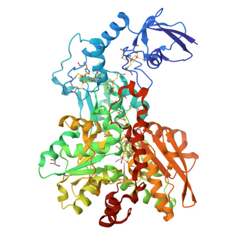
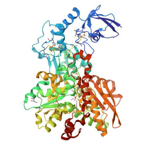
1.39 Angstrom crystal structure of Fe-only hydrogenase


Experimental Data Snapshot

  • Method: X-RAY DIFFRACTION
  • Resolution: 1.39 Å
  • R-Value Free: 0.194 
  • R-Value Work: 0.132 

Starting Model: experimental
View more details

wwPDB Validation   3D Report Full Report


Ligand Structure Quality Assessment 


This is version 1.2 of the entry. See complete history


Literature

Dithiomethylether as a ligand in the hydrogenase h-cluster.

Pandey, A.S.Harris, T.V.Giles, L.J.Peters, J.W.Szilagyi, R.K.

(2008) J Am Chem Soc 130: 4533-4540

  • DOI: https://doi.org/10.1021/ja711187e
  • Primary Citation of Related Structures:  
    3C8Y

  • PubMed Abstract: 

    An X-ray crystallographic refinement of the H-cluster of [FeFe]-hydrogenase from Clostridium pasteurianum has been carried out to close-to atomic resolution and is the highest resolution [FeFe]-hydrogenase presented to date. The 1.39 A, anisotropically refined [FeFe]-hydrogenase structure provides a basis for examining the outstanding issue of the composition of the unique nonprotein dithiolate ligand of the H-cluster. In addition to influencing the electronic structure of the H-cluster, the composition of the ligand has mechanistic implications due to the potential of the bridge-head gamma-group participating in proton transfer during catalysis. In this work, sequential density functional theory optimizations of the dithiolate ligand embedded in a 3.5-3.9 A protein environment provide an unbiased approach to examining the most likely composition of the ligand. Structural, conformational, and energetic considerations indicate a preference for dithiomethylether as an H-cluster ligand and strongly disfavor the dithiomethylammonium as a catalytic base for hydrogen production.


  • Organizational Affiliation

    Department of Chemistry and Biochemistry and Astrobiology Biogeocatalysis Research Center, Montana State University, Bozeman, Montana 59717, USA.


Macromolecules
Find similar proteins by:  (by identity cutoff)  |  3D Structure
Entity ID: 1
MoleculeChains Sequence LengthOrganismDetailsImage
Iron hydrogenase 1574Clostridium pasteurianumMutation(s): 0 
EC: 1.12.7.2
UniProt
Find proteins for P29166 (Clostridium pasteurianum)
Go to UniProtKB:  P29166
Sequence Annotations
Expand
  • Reference Sequence
Small Molecules
Ligands 4 Unique
IDChains Name / Formula / InChI Key2D Diagram3D Interactions
HCN
Query on HCN

Download Ideal Coordinates CCD File 
B [auth A]2 IRON/2 SULFUR/3 CARBONYL/2 CYANIDE/WATER/METHYLETHER CLUSTER
C7 H10 Fe2 N2 O5 S2
GIQCNDUJZDHMQH-UHFFFAOYSA-N
SF4
Query on SF4

Download Ideal Coordinates CCD File 
C [auth A],
D [auth A],
E [auth A],
F [auth A]
IRON/SULFUR CLUSTER
Fe4 S4
LJBDFODJNLIPKO-UHFFFAOYSA-N
FES
Query on FES

Download Ideal Coordinates CCD File 
G [auth A]FE2/S2 (INORGANIC) CLUSTER
Fe2 S2
NIXDOXVAJZFRNF-UHFFFAOYSA-N
GOL
Query on GOL

Download Ideal Coordinates CCD File 
H [auth A],
I [auth A],
J [auth A]
GLYCEROL
C3 H8 O3
PEDCQBHIVMGVHV-UHFFFAOYSA-N
Experimental Data & Validation

Experimental Data

  • Method: X-RAY DIFFRACTION
  • Resolution: 1.39 Å
  • R-Value Free: 0.194 
  • R-Value Work: 0.132 
  • Space Group: P 42 21 2
Unit Cell:
Length ( Å )Angle ( ˚ )
a = 110.75α = 90
b = 110.75β = 90
c = 103.54γ = 90
Software Package:
Software NamePurpose
MOSFLMdata reduction
SCALAdata scaling
MOLREPphasing
SHELXrefinement
PDB_EXTRACTdata extraction
SHELXL-97refinement

Structure Validation

View Full Validation Report


Ligand Structure Quality Assessment 


Entry History 

Deposition Data

Revision History  (Full details and data files)

  • Version 1.0: 2008-04-22
    Type: Initial release
  • Version 1.1: 2011-07-13
    Changes: Non-polymer description, Version format compliance
  • Version 1.2: 2023-08-30
    Changes: Data collection, Database references, Derived calculations, Refinement description