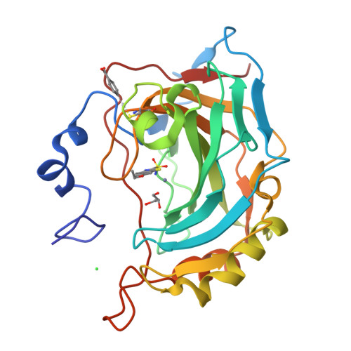
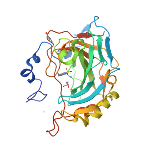
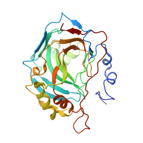
3CAJ

Crystal structure of the human carbonic anhydrase II in complex with ethoxzolamide


Experimental Data Snapshot

  • Method: X-RAY DIFFRACTION
  • Resolution: 1.80 Å
  • R-Value Free: 0.214 
  • R-Value Work: 0.180 

Starting Model: experimental
View more details

wwPDB Validation   3D Report Full Report


Ligand Structure Quality Assessment 


This is version 1.3 of the entry. See complete history


Literature

Carbonic anhydrase inhibitors: the X-ray crystal structure of ethoxzolamide complexed to human isoform II reveals the importance of thr200 and gln92 for obtaining tight-binding inhibitors

Di Fiore, A.Pedone, C.Antel, J.Waldeck, H.Witte, A.Wurl, M.Scozzafava, A.Supuran, C.T.De Simone, G.

(2008) Bioorg Med Chem Lett 18: 2669-2674

  • DOI: https://doi.org/10.1016/j.bmcl.2008.03.023
  • Primary Citation of Related Structures:  
    3CAJ

  • PubMed Abstract: 

    Ethoxzolamide, an almost forgotten inhibitor of the metalloenzyme carbonic anhydrase (CA, EC 4.2.1.1), is the only classical inhibitor whose structure in adduct with any isoform was not reported yet. We report here the inhibition data of this molecule with the 12 catalytically active mammalian isozymes (CA I-CA XIV) and the X-ray crystal structure with the cytosolic, ubiquitous isoform CA II. These data are presumably useful for the design of novel CA inhibitors, targeting various CA isozymes, considering that ethoxzolamide was already the lead molecule to obtain the second generation inhibitors, dorzolamide and brinzolamide, clinically used antiglaucoma agents with topical action, as well as various other investigational agents.


  • Organizational Affiliation

    Istituto di Biostrutture e Bioimmagini-CNR, via Mezzocannone 16, 80134 Napoli, Italy.


Macromolecules
Find similar proteins by:  (by identity cutoff)  |  3D Structure
Entity ID: 1
MoleculeChains Sequence LengthOrganismDetailsImage
Carbonic anhydrase 2260Homo sapiensMutation(s): 0 
EC: 4.2.1.1 (PDB Primary Data), 4.2.1.69 (UniProt)
UniProt & NIH Common Fund Data Resources
Find proteins for P00918 (Homo sapiens)
Explore P00918 
Go to UniProtKB:  P00918
PHAROS:  P00918
GTEx:  ENSG00000104267 
Entity Groups  
Sequence Clusters30% Identity50% Identity70% Identity90% Identity95% Identity100% Identity
UniProt GroupP00918
Sequence Annotations
Expand
  • Reference Sequence
Small Molecules
Binding Affinity Annotations 
IDSourceBinding Affinity
EZL PDBBind:  3CAJ Ki: 8 (nM) from 1 assay(s)
Experimental Data & Validation

Experimental Data

  • Method: X-RAY DIFFRACTION
  • Resolution: 1.80 Å
  • R-Value Free: 0.214 
  • R-Value Work: 0.180 
  • Space Group: P 1 21 1
Unit Cell:
Length ( Å )Angle ( ˚ )
a = 42.21α = 90
b = 41.36β = 104.35
c = 72.03γ = 90
Software Package:
Software NamePurpose
MAR345data collection
CNSrefinement
DENZOdata reduction
SCALEPACKdata scaling
CNSphasing

Structure Validation

View Full Validation Report


Ligand Structure Quality Assessment 


Entry History 

Deposition Data

Revision History  (Full details and data files)

  • Version 1.0: 2008-04-08
    Type: Initial release
  • Version 1.1: 2011-07-13
    Changes: Non-polymer description, Version format compliance
  • Version 1.2: 2017-10-25
    Changes: Refinement description
  • Version 1.3: 2023-11-01
    Changes: Data collection, Database references, Derived calculations, Refinement description