3FHB

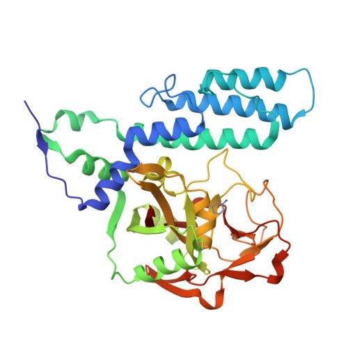
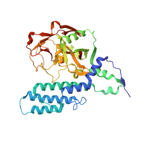
Human poly(ADP-ribose) polymerase 3, catalytic fragment in complex with an inhibitor 3-aminobenzoic acid


Experimental Data Snapshot

  • Method: X-RAY DIFFRACTION
  • Resolution: 2.30 Å
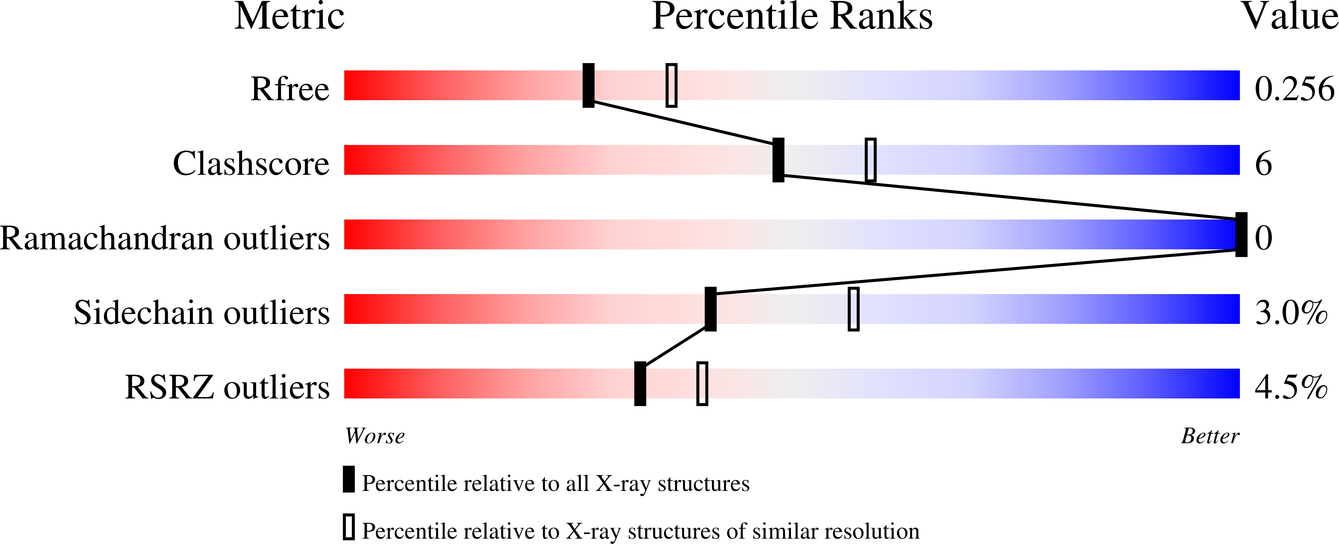
  • R-Value Free: 0.254 
  • R-Value Work: 0.178 
  • R-Value Observed: 0.182 

wwPDB Validation   3D Report Full Report


This is version 1.3 of the entry. See complete history


Literature

Structural basis for inhibitor specificity in human poly(ADP-ribose) polymerase-3.

Lehtio, L.Jemth, A.S.Collins, R.Loseva, O.Johansson, A.Markova, N.Hammarstrom, M.Flores, A.Holmberg-Schiavone, L.Weigelt, J.Helleday, T.Schuler, H.Karlberg, T.

(2009) J Med Chem 52: 3108-3111

  • DOI: https://doi.org/10.1021/jm900052j
  • Primary Citation of Related Structures:  
    3C49, 3C4H, 3CE0, 3FHB

  • PubMed Abstract: 

    Poly(ADP-ribose) polymerases (PARPs) activate DNA repair mechanisms upon stress- and cytotoxin-induced DNA damage, and inhibition of PARP activity is a lead in cancer drug therapy. We present a structural and functional analysis of the PARP domain of human PARP-3 in complex with several inhibitors. Of these, KU0058948 is the strongest inhibitor of PARP-3 activity. The presented crystal structures highlight key features for potent inhibitor binding and suggest routes for creating isoenzyme-specific PARP inhibitors.


  • Organizational Affiliation

    Structural Genomics Consortium, Department of Medical Biochemistry and Biophysics, Karolinska Institutet, SE-17177 Stockholm, Sweden.


Macromolecules
Find similar proteins by:  (by identity cutoff)  |  3D Structure
Entity ID: 1
MoleculeChains Sequence LengthOrganismDetailsImage
Poly [ADP-ribose] polymerase 3357Homo sapiensMutation(s): 0 
Gene Names: PARP3ADPRT3ADPRTL3
EC: 2.4.2.30 (PDB Primary Data), 2.4.2 (UniProt)
UniProt & NIH Common Fund Data Resources
Find proteins for Q9Y6F1 (Homo sapiens)
Explore Q9Y6F1 
Go to UniProtKB:  Q9Y6F1
PHAROS:  Q9Y6F1
GTEx:  ENSG00000041880 
Entity Groups  
Sequence Clusters30% Identity50% Identity70% Identity90% Identity95% Identity100% Identity
UniProt GroupQ9Y6F1
Sequence Annotations
Expand
  • Reference Sequence
Small Molecules
Ligands 1 Unique
IDChains Name / Formula / InChI Key2D Diagram3D Interactions
GAB
Query on GAB

Download Ideal Coordinates CCD File 
B [auth A]3-AMINOBENZOIC ACID
C7 H7 N O2
XFDUHJPVQKIXHO-UHFFFAOYSA-N
Binding Affinity Annotations 
IDSourceBinding Affinity
GAB PDBBind:  3FHB Kd: 2000 (nM) from 1 assay(s)
BindingDB:  3FHB Kd: 2000 (nM) from 1 assay(s)
Experimental Data & Validation

Experimental Data

  • Method: X-RAY DIFFRACTION
  • Resolution: 2.30 Å
  • R-Value Free: 0.254 
  • R-Value Work: 0.178 
  • R-Value Observed: 0.182 
  • Space Group: P 1 21 1
Unit Cell:
Length ( Å )Angle ( ˚ )
a = 55.24α = 90
b = 56.7β = 112.86
c = 56.48γ = 90
Software Package:
Software NamePurpose
ProDCdata collection
PHASERphasing
REFMACrefinement
XDSdata reduction
XSCALEdata scaling

Structure Validation

View Full Validation Report


Entry History 

Revision History  (Full details and data files)

  • Version 1.0: 2009-01-06
    Type: Initial release
  • Version 1.1: 2011-07-13
    Changes: Version format compliance
  • Version 1.2: 2017-11-01
    Changes: Refinement description
  • Version 1.3: 2024-02-21
    Changes: Data collection, Database references, Derived calculations