3NBS | pdb_00003nbs

Crystal structure of dimeric cytochrome c from horse heart


Experimental Data Snapshot

  • Method: X-RAY DIFFRACTION
  • Resolution: 2.20 Å
  • R-Value Free: 
    0.273 (Depositor), 0.270 (DCC) 
  • R-Value Work: 
    0.215 (Depositor), 0.210 (DCC) 
  • R-Value Observed: 
    0.215 (Depositor) 

Starting Model: experimental
View more details

wwPDB Validation   3D Report Full Report


Ligand Structure Quality Assessment 

Created with Raphaël 2.3.0Worse 01 BetterLigand structure goodness of fit to experimental dataBest fitted HECClick on this verticalbar to view details

This is version 2.2 of the entry. See complete history


Literature

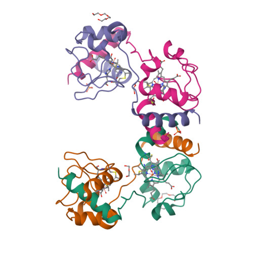
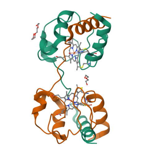
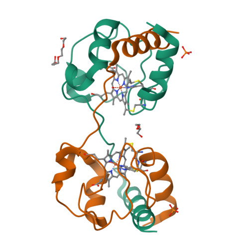
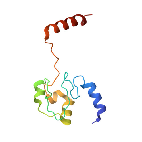
Cytochrome c polymerization by successive domain swapping at the C-terminal helix

Hirota, S.Hattori, Y.Nagao, S.Taketa, M.Komori, H.Kamikubo, H.Wang, Z.Takahashi, I.Negi, S.Sugiura, Y.Kataoka, M.Higuchi, Y.

(2010) Proc Natl Acad Sci U S A 107: 12854-12859

  • DOI: https://doi.org/10.1073/pnas.1001839107
  • Primary Citation of Related Structures:  
    3NBS, 3NBT

  • PubMed Abstract: 

    Cytochrome c (cyt c) is a stable protein that functions in a monomeric state as an electron donor for cytochrome c oxidase. It is also released to the cytosol when permeabilization of the mitochondrial outer membrane occurs at the early stage of apoptosis. For nearly half a century, it has been known that cyt c forms polymers, but the polymerization mechanism remains unknown. We found that cyt c forms polymers by successive domain swapping, where the C-terminal helix is displaced from its original position in the monomer and Met-heme coordination is perturbed significantly. In the crystal structures of dimeric and trimeric cyt c, the C-terminal helices are replaced by the corresponding domain of other cyt c molecules and Met80 is dissociated from the heme. The solution structures of dimeric, trimeric, and tetrameric cyt c were linear based on small-angle X-ray scattering measurements, where the trimeric linear structure shifted toward the cyclic structure by addition of PEG and (NH(4))(2)HPO(4). The absorption and CD spectra of high-order oligomers (approximately 40 mer) were similar to those of dimeric and trimeric cyt c but different from those of monomeric cyt c. For dimeric, trimeric, and tetrameric cyt c, the DeltaH of the oligomer dissociation to monomers was estimated to be about -20 kcal/mol per protomer unit, where Met-heme coordination appears to contribute largely to DeltaH. The present results suggest that cyt c polymerization occurs by successive domain swapping, which may be a common mechanism of protein polymerization.


  • Organizational Affiliation

    Graduate School of Materials Science, Nara Institute of Science and Technology, 8916-5 Takayama, Ikoma, Nara 630-0192, Japan. hirota@ms.naist.jp


Macromolecules
Find similar proteins by:  (by identity cutoff)  |  3D Structure
Entity ID: 1
MoleculeChains Sequence LengthOrganismDetailsImage
Cytochrome c
A, B, C, D
104Equus caballusMutation(s): 0 
UniProt
Find proteins for P00004 (Equus caballus)
Explore P00004 
Go to UniProtKB:  P00004
Entity Groups  
Sequence Clusters30% Identity50% Identity70% Identity90% Identity95% Identity100% Identity
UniProt GroupP00004
Sequence Annotations
Expand
  • Reference Sequence
Small Molecules
Ligands 4 Unique
IDChains Name / Formula / InChI Key2D Diagram3D Interactions
HEC
Query on HEC

Download Ideal Coordinates CCD File 
E [auth A],
G [auth B],
I [auth C],
L [auth D]
HEME C
C34 H34 Fe N4 O4
HXQIYSLZKNYNMH-LJNAALQVSA-N
PG4
Query on PG4

Download Ideal Coordinates CCD File 
F [auth A],
K [auth C]
TETRAETHYLENE GLYCOL
C8 H18 O5
UWHCKJMYHZGTIT-UHFFFAOYSA-N
PEG
Query on PEG

Download Ideal Coordinates CCD File 
H [auth B],
N [auth D]
DI(HYDROXYETHYL)ETHER
C4 H10 O3
MTHSVFCYNBDYFN-UHFFFAOYSA-N
PO4
Query on PO4

Download Ideal Coordinates CCD File 
J [auth C],
M [auth D]
PHOSPHATE ION
O4 P
NBIIXXVUZAFLBC-UHFFFAOYSA-K
Experimental Data & Validation

Experimental Data

  • Method: X-RAY DIFFRACTION
  • Resolution: 2.20 Å
  • R-Value Free:  0.273 (Depositor), 0.270 (DCC) 
  • R-Value Work:  0.215 (Depositor), 0.210 (DCC) 
  • R-Value Observed: 0.215 (Depositor) 
Space Group: P 1
Unit Cell:
Length ( Å )Angle ( ˚ )
a = 41.753α = 66.28
b = 56.305β = 89.94
c = 60.815γ = 73.66
Software Package:
Software NamePurpose
HKL-2000data collection
MOLREPphasing
CNSrefinement
HKL-2000data reduction
HKL-2000data scaling

Structure Validation

View Full Validation Report


Ligand Structure Quality Assessment 

Created with Raphaël 2.3.0Worse 01 BetterLigand structure goodness of fit to experimental dataBest fitted HECClick on this verticalbar to view details

Entry History 

Deposition Data

Revision History  (Full details and data files)

  • Version 1.0: 2010-07-14
    Type: Initial release
  • Version 1.1: 2011-07-13
    Changes: Version format compliance
  • Version 1.2: 2017-11-08
    Changes: Refinement description
  • Version 2.0: 2019-10-02
    Changes: Advisory, Atomic model, Data collection, Derived calculations, Non-polymer description, Structure summary
  • Version 2.1: 2023-11-01
    Changes: Data collection, Database references, Derived calculations, Refinement description
  • Version 2.2: 2024-10-16
    Changes: Structure summary