3TD1 | pdb_00003td1

Crystal structure of the bacterial A1408G-mutant and the protozoa cytoplasmic ribosomal decoding site in complex with geneticin

  • Classification: RNA/ANTIBIOTIC
  • Mutation(s): No 

  • Deposited: 2011-08-10 Released: 2011-12-07 
  • Deposition Author(s): Kondo, J.

Experimental Data Snapshot

  • Method: X-RAY DIFFRACTION
  • Resolution: 2.10 Å
  • R-Value Free: 
    0.243 (Depositor), 0.240 (DCC) 
  • R-Value Work: 
    0.196 (Depositor), 0.180 (DCC) 

Starting Model: experimental
View more details

wwPDB Validation   3D Report Full Report


Ligand Structure Quality Assessment 

Created with Raphaël 2.3.0Worse 01 BetterLigand structure goodness of fit to experimental dataBest fitted GETClick on this verticalbar to view details

This is version 1.2 of the entry. See complete history


Literature

A structural basis for the antibiotic resistance conferred by an A1408G mutation in 16S rRNA and for the antiprotozoal activity of aminoglycosides

Kondo, J.

(2012) Angew Chem Int Ed Engl 51: 465-468

  • DOI: https://doi.org/10.1002/anie.201106084
  • Primary Citation of Related Structures:  
    3TD0, 3TD1

  • PubMed Abstract: 

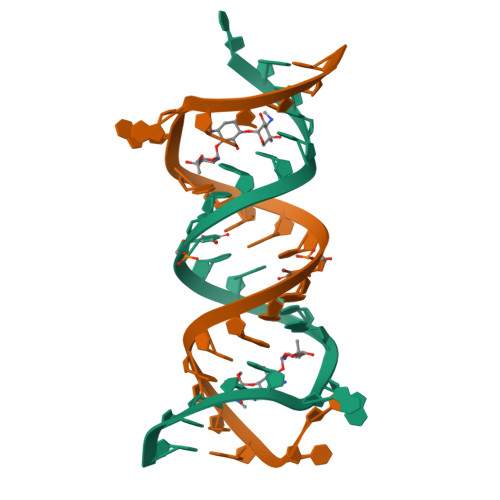
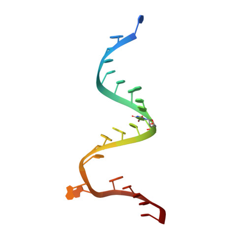
    Resistance explained: The crystal structures of the ribosomal decoding A site with an A1408G antibiotic-resistance mutation were solved in the presence and absence of the aminoglycoside geneticin (see structure, geneticin carbon framework in yellow). These structures show how bacteria acquire high-level resistance against aminoglycosides by the mutation.


  • Organizational Affiliation

    Department of Materials and Life Sciences, Faculty of Science and Technology, Sophia University, 7-1 Kioi-cho, Chiyoda-ku, 102-8554 Tokyo, Japan. j.kondo@sophia.ac.jp


Macromolecules

Find similar nucleic acids by:  Sequence   |   3D Structure  

Entity ID: 1
MoleculeChains LengthOrganismImage
RNA (5'-R(*UP*UP*GP*CP*GP*UP*CP*GP*CP*GP*(5BU)P*CP*GP*AP*CP*GP*AP*AP*GP*UP*CP*GP*C)-3')
A, B
23N/A
Sequence Annotations
Expand
  • Reference Sequence
Experimental Data & Validation

Experimental Data

  • Method: X-RAY DIFFRACTION
  • Resolution: 2.10 Å
  • R-Value Free:  0.243 (Depositor), 0.240 (DCC) 
  • R-Value Work:  0.196 (Depositor), 0.180 (DCC) 
Space Group: P 21 21 2
Unit Cell:
Length ( Å )Angle ( ˚ )
a = 33.47α = 90
b = 88.3β = 90
c = 46.21γ = 90
Software Package:
Software NamePurpose
PHENIXmodel building
CNSrefinement
d*TREKdata reduction
SCALAdata scaling
PHENIXphasing

Structure Validation

View Full Validation Report


Ligand Structure Quality Assessment 

Created with Raphaël 2.3.0Worse 01 BetterLigand structure goodness of fit to experimental dataBest fitted GETClick on this verticalbar to view details

Entry History 

Deposition Data

  • Released Date: 2011-12-07 
  • Deposition Author(s): Kondo, J.

Revision History  (Full details and data files)

  • Version 1.0: 2011-12-07
    Type: Initial release
  • Version 1.1: 2014-03-12
    Changes: Database references
  • Version 1.2: 2023-11-01
    Changes: Data collection, Database references, Derived calculations, Refinement description